Bionic bone tissue engineering bracket as well as preparation method and application thereof

A tissue engineering scaffold, bionic bone technology, used in tissue regeneration, pharmaceutical formulations, coatings, etc.

Active Publication Date: 2017-10-20
SOUTH CHINA NORMAL UNIVERSITY
View PDF9 Cites 2 Cited by
  • Summary
  • Abstract
  • Description
  • Claims
  • Application Information

AI Technical Summary

Problems solved by technology

[0004] Although various strategies for modifying scaffolds for bone tissue engineerin

Method used


Image

  • Bionic bone tissue engineering bracket as well as preparation method and application thereof
  • Bionic bone tissue engineering bracket as well as preparation method and application thereof
  • Bionic bone tissue engineering bracket as well as preparation method and application thereof

Examples


Embodiment 1

[0084] Example 1 Preparation of Biomimetic Bone Tissue Engineering Scaffold AG-o-CNTs

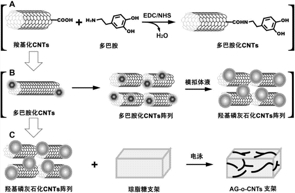
[0085] The preparation process of the bionic bone tissue engineering scaffold of the present invention is as follows: figure 1 As shown, first, carboxylated multi-walled carbon nanotubes (COOH-CNTs) are modified by dopamine to form dopamine-coated multi-walled carbon nanotubes (Dopamine-CNTs, D-CNTs). Afterwards, place this type of carbon nanotubes in simulated body fluid for 4-8 weeks, and the phosphorus ions and calcium ions in the simulated body fluid will form hydroxyapatite under the guidance of phenolic hydroxyl groups on dopamine. Walled carbon nanotubes (Hydroxyapatite-CNTS, H-CNTs, that is, multi-walled carbon nanotubes loaded with hydroxyapatite). Afterwards, the H-CNTs were formed into an ordered parallel array in the agarose gel by electrophoresis, thereby constructing a biomimetic ordered agarose-carbon nanotube scaffold (AG-o-CNTs scaffold).

[0086] Specifically, the prepa...

Embodiment 2

[0105] Example 2 Characterization of AG-o-CNTs Biomimetic Bone Tissue Engineering Scaffold

[0106] 1. Infrared spectrum detection

[0107] (1) Dry each sample and KBr in a dryer, mix 1-2 mg of sample with 200 mg of pure KBr and grind them evenly, and grind the mixture to a particle size of less than 2 μm to avoid the influence of scattered light. Put the mixture in the mold, press the mixture into a transparent sheet with a pressure of 5-10MPa on the hydraulic press, and wait for the machine to be determined; at the same time, dry the blank bracket and the modified bracket, and test it on the machine.

[0108] (2) Infrared detection of CNTs after dopamine and hydroxyapatite modification

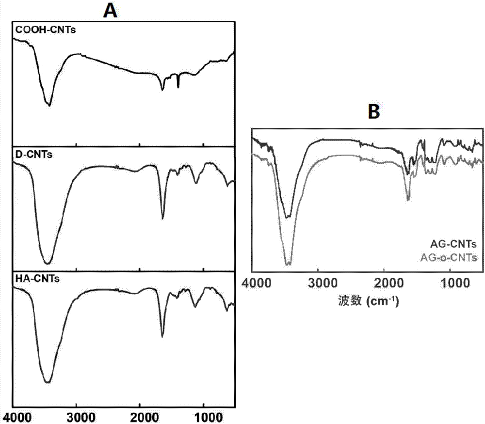
[0109] To further determine the composition of the structures generated on the surface of sh-CNTs, we first analyzed the samples by Fourier transform infrared spectroscopy detection.

[0110] experiment such as figure 2 As shown in Figure A, the results show that the infrared characteris...

Embodiment 3

[0137] Example 3 Application of Biomimetic Bone Tissue Engineering Scaffold AG-o-CNTs

[0138] The role of AG-o-CNTs biomimetic scaffold in promoting the growth of MSCs was studied, and the carbon nanotube modified agarose scaffold (AG-CNTs scaffold) without electrophoresis treatment was used as a control.

[0139] 1. Cell culture

[0140] Place the blank and modified scaffolds in 24-well cell culture plates 1×1×1cm 3 , after the cells were cultured to 60-90% confluence in the culture flask, they were treated with 1×10 4 -3×10 4 The density per well was inoculated onto 24-well plates and cultured for 2, 4, and 6 days for subsequent experiments. Other cell culture conditions are: low-sugar DMEM medium containing 10% newborn calf serum, 37°C, 5.0% CO 2 .

[0141] In order to study the effect of the two scaffold materials on cell growth, we seeded rat bone mesenchymal stem cells (bMSCs) on the scaffolds and detected their growth status.

[0142] 2. Immunofluorescence (DAPI)...


PUM


Abstract

The invention discloses a bionic bone tissue engineering bracket as well as a preparation method and application thereof. The method comprises the following steps: firstly, performing carboxylation and dopamine surface modification on carbon nano tubes (CNTs) in turn; forming hydroxyapatite grains on the dopamine CNTs surfaces under the condition of simulated body fluid, thereby constructing biological mineralized CNTs; utilizing agarose to construct a main body bracket and utilizing an electrophoretic technique to move the mineralized CNTs in the bracket and form an array in parallel in sequence, thereby constructing the bionic bone tissue engineering bracket AG-o-CNTs. The bionic bone tissue engineering bracket AG-o-CNTs disclosed by the invention can effectively promote the growth of mesenchymal stem cells (bMSCs) and has wide application prospect at the aspect of bone tissue engineering bracket material or bone substitute material.

Description

technical field [0001] The invention belongs to the technical field of biomedical materials. More specifically, it relates to a biomimetic bone tissue engineering scaffold based on agarose gel and ordered multi-walled carbon nanotubes and its preparation method and application. Background technique [0002] The importance of bone tissue and the harm caused by bone-related diseases have accelerated the development of bone tissue engineering. In this context, bone tissue engineering has become a new frontier method for repairing damaged organs because of its great potential to treat bone-related injuries or diseases. Generally speaking, the research on bone tissue engineering includes three major elements: scaffolds, cells and growth factors. Among the above three, the research on stents is one of the more active research fields at present. Bone tissue engineering requires that the scaffold used is not only an ideal bone substitute, but also has the ability to induce bone c...

Claims


Application Information

Patent Timeline
IPC IPC(8): A61L27/20A61L27/08A61L27/28A61L27/32A61L27/38
CPCA61L27/08A61L27/20A61L27/28A61L27/32A61L27/3834A61L27/3847A61L2400/18A61L2430/02C08L5/12
Inventor 关燕清陈吾雅
Owner SOUTH CHINA NORMAL UNIVERSITY
Why Eureka
Try Eureka