A bionic bone tissue engineering scaffold and its preparation method and application
A tissue engineering scaffold, bionic bone technology, used in tissue regeneration, pharmaceutical formulations, prostheses, etc.
- Summary
- Abstract
- Description
- Claims
- Application Information
AI Technical Summary
Problems solved by technology
Method used
Image
Examples
Embodiment 1
[0084] Example 1 Preparation of Biomimetic Bone Tissue Engineering Scaffold AG-o-CNTs
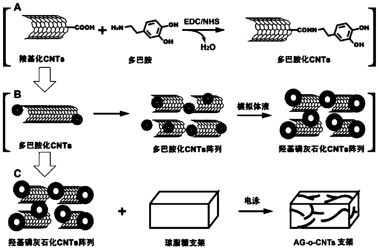
[0085] The preparation process of the bionic bone tissue engineering scaffold of the present invention is as follows: figure 1 As shown, first, carboxylated multi-walled carbon nanotubes (COOH-CNTs) are modified by dopamine to form dopamine-coated multi-walled carbon nanotubes (Dopamine-CNTs, D-CNTs). Afterwards, place this type of carbon nanotubes in simulated body fluid for 4-8 weeks, and the phosphorus ions and calcium ions in the simulated body fluid will form hydroxyapatite under the guidance of phenolic hydroxyl groups on dopamine. Walled carbon nanotubes (Hydroxyapatite-CNTS, H-CNTs, that is, multi-walled carbon nanotubes loaded with hydroxyapatite). Afterwards, the H-CNTs were formed into an ordered parallel array in the agarose gel by electrophoresis, thereby constructing a biomimetic ordered agarose-carbon nanotube scaffold (AG-o-CNTs scaffold).
[0086] Specifically, the prepa...
Embodiment 2
[0105] Example 2 Characterization of AG-o-CNTs Biomimetic Bone Tissue Engineering Scaffold
[0106] 1. Infrared spectrum detection
[0107] (1) Dry each sample and KBr in a dryer, mix 1-2 mg of sample with 200 mg of pure KBr and grind them evenly, and grind the mixture to a particle size of less than 2 μm to avoid the influence of scattered light. Put the mixture in the mold, press the mixture into a transparent sheet with a pressure of 5-10MPa on the hydraulic press, and wait for the machine to be determined; at the same time, dry the blank bracket and the modified bracket, and test it on the machine.
[0108] (2) Infrared detection of CNTs after dopamine and hydroxyapatite modification
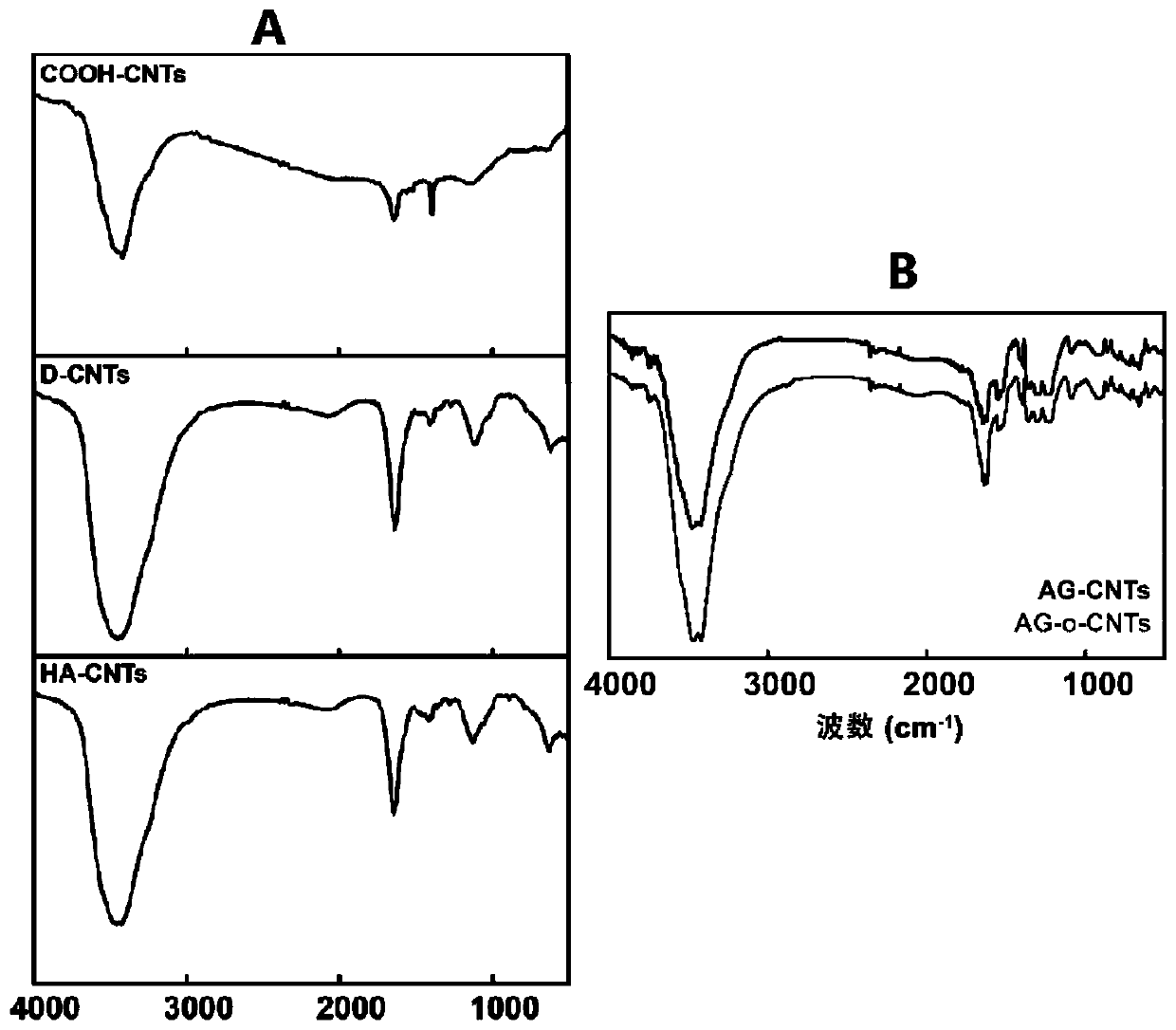
[0109] To further determine the composition of the structures generated on the surface of sh-CNTs, we first analyzed the samples by Fourier transform infrared spectroscopy detection.
[0110] experiment as figure 2 As shown in Figure A, the results show that the infrared characteristic p...
Embodiment 3
[0137] Example 3 Application of bionic bone tissue engineering scaffold AG-o-CNTs
[0138] To study the effect of AG-o-CNTs biomimetic scaffold in promoting the growth of MSCs, and use the carbon nanotube modified agarose scaffold (AG-CNTs scaffold) without electrophoresis as a control.
[0139] 1. Cell culture
[0140] Place blank and modified scaffolds 1×1×1cm in 24-well cell culture plates respectively 3 , the cells were cultured to 60-90% confluency in the 4 -3×10 4 The density of / well was seeded into 24-well plates and cultured for 2, 4, and 6 days for subsequent experiments. Other cell culture conditions were: low-glucose DMEM medium containing 10% newborn calf serum, 37°C, 5.0% CO 2 .
[0141] To study the effect of the two scaffold materials on cell growth, we seeded rat bone mesenchymal stem cells (bMSCs) on the scaffolds and examined their growth status.
[0142] 2. Immunofluorescence (DAPI) detection
[0143] Firstly, the density and growth distribution of c...
PUM
| Property | Measurement | Unit |
|---|---|---|
| diameter | aaaaa | aaaaa |
| diameter | aaaaa | aaaaa |
| diameter | aaaaa | aaaaa |
Abstract
Description
Claims
Application Information
 - R&D
- Intellectual Property
- Life Sciences
- Materials
- Tech Scout
- Unparalleled Data Quality
- Higher Quality Content
- 60% Fewer Hallucinations
Browse by: Latest US Patents, China's latest patents, Technical Efficacy Thesaurus, Application Domain, Technology Topic, Popular Technical Reports.
© 2025 PatSnap. All rights reserved.Legal|Privacy policy|Modern Slavery Act Transparency Statement|Sitemap|About US| Contact US: help@patsnap.com


