
Culture method for fibroblast derived from autologous body skin

A technology of fibroblasts and culture methods, which is applied in the field of autologous skin-derived fibroblasts culture, can solve the problems of low fibroblast purity, large influence of miscellaneous cells, and large fibroblast damage, so as to improve cell yield, The effect of fast cell proliferation and improved purity

Active Publication Date: 2017-11-17
SHANGHAI BIOMED UNION BIOTECHNOLOGY CO LTD
View PDF4 Cites 11 Cited by
  • Summary
  • Abstract
  • Description
  • Claims
  • Application Information

AI Technical Summary

Problems solved by technology

However, the use of enzyme digestion to remove the epidermis in the early stage will cause greater damage to fibroblasts, and the medium of this method has no selectivity for fibroblasts, and the impact of miscellaneous cells is relatively large. The purity of the cultured primary fibroblasts is low and needs to be subcultured. Fibroblasts with high purity can be obtained several times

Method used


Image

  • Culture method for fibroblast derived from autologous body skin
  • Culture method for fibroblast derived from autologous body skin
  • Culture method for fibroblast derived from autologous body skin

Examples


Embodiment 1

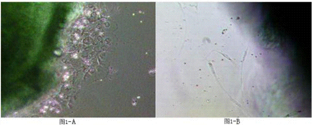
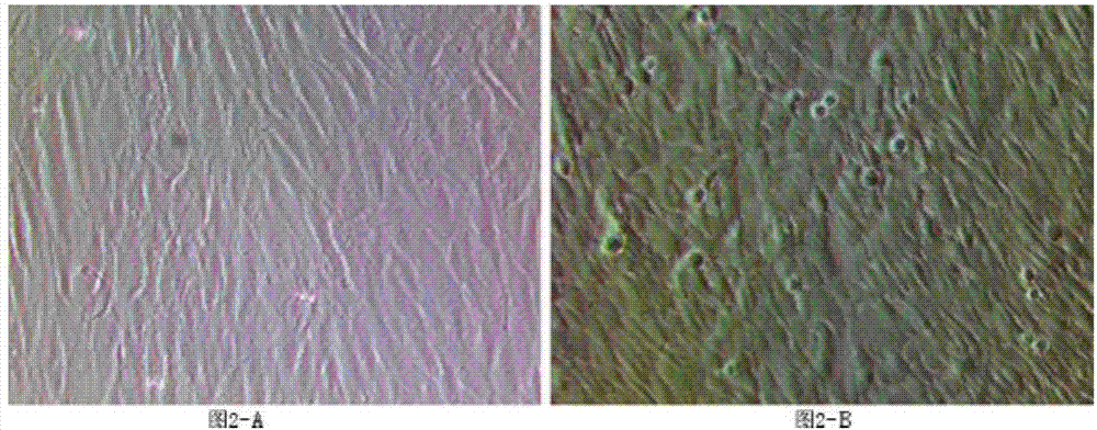
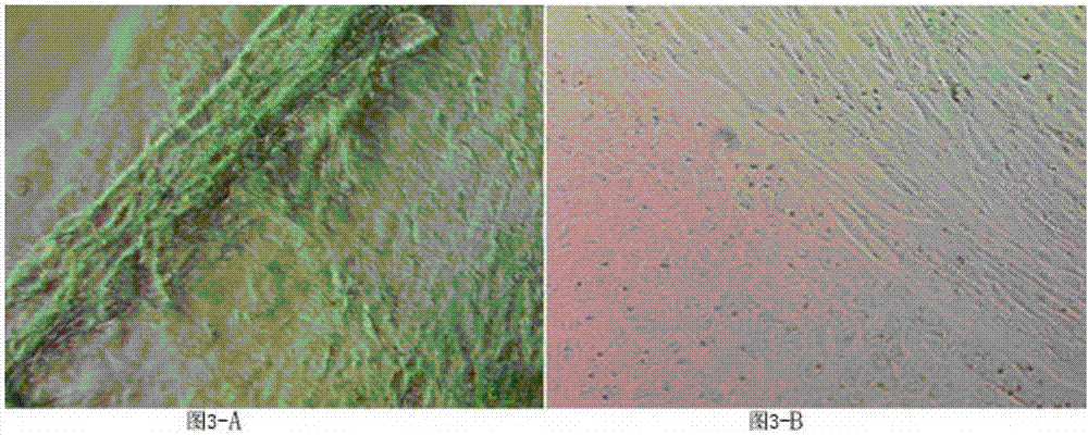
[0040] A kind of fibroblast culture method derived from autologous skin

[0041] test group

[0042] 1) Configure selection medium:

[0043] Mix DMEM medium and F12 medium at a volume ratio of 3-1:1 to form Xml basal medium, then add (2ng-9ng)*Xml b-FGF, (1μg-20μg) to the basal medium *Xml of IGF-1 (Insulin-likeGrowthFactors, insulin-like No. 1 growth factor), (0.1 μg-1μg) *Xml of EGF, then add the above-mentioned medium and fetal bovine serum according to the volume ratio of each cytokine 4: 1 Prepare a selective medium to accelerate the proliferation rate of fibroblasts and inhibit the growth of other cells.

[0044]Among them, DMEM medium and F12 medium are mixed as a serum-free basal medium, and the advantages of various components in F12 medium and high-concentration nutrients in DMEM medium are used to make fibroblasts proliferate quickly and require serum volume was significantly reduced. DMEM medium and F12 medium are preferably mixed according to the volume ratio ...

Embodiment 2

[0073] Verification experiment A

[0074] Fluorescence staining experiment of second generation fibroblasts

[0075] In order to verify and compare the purity of the fibroblasts cultured in the experimental group and the control group in Example 1, a fibroblast fluorescent staining experiment was performed to compare the effects of the two methods.

[0076] Because the fibroblast culture method of the experimental group has fibroblast selectivity, the primary fibroblasts cultured by this method have a very high purity, while the fibroblasts cultured according to the culture method of the control group grow with passage. Purification, so the second-generation fibroblasts were selected for experiments to compare the culture effects of the two groups of culture methods, so as to avoid excessive passage and lead to no difference in the purity of the two groups of cells.

[0077] 1) Take the culture medium of the second-generation fibroblasts of the experimental group and the cont...

Embodiment 3

[0086] Verification experiment B


PUM


Abstract

The invention discloses a culture method for fibroblast derived from autologous body skin. The method comprises the following steps: 1) taking autologous body skin, dividing the autologous body skin into multiple tissue blocks, attaching the tissue blocks to a culture dish, dropwise adding concentrated feta calf serum on the tissue blocks, completely soaking the tissue blocks, preventing the tissue blocks from drying and promoting the fibroblast to crawl out; 2) adding a selective culture medium into the culture dish in the step 1), placing the culture dish into an incubator to culture until the fibroblast fusion is finished, wherein the selective culture medium is used for promoting the fibroblast to grow and inhibiting other parenchyma cells to grow; 3) taking the culture dish in the step 2) out of the incubator, adding pancreatin for digestion to enable the fibroblast to fall off from the culture dish; 4) inoculating the fibroblast obtained in the step 3) into a culture bottle by a complete culture medium, enabling the fibroblast density to reach 90 percent to obtain primary fibroblast, wherein the complete culture medium is used for ending the digestion in the step 3) and is used as a culture medium for growth and proliferation of the fibroblast; 5) passaging the bottle-expanding culture of the primary fibroblast in the step 4).

Description

technical field [0001] The invention relates to the field of cell culture, in particular to a method for culturing fibroblasts derived from autologous skin. Background technique [0002] Skin fibroblasts are widely distributed in the dermis. Fibroblasts secrete a variety of substances to form the extracellular matrix, thereby forming connective tissue and exerting structural functions on the body. Fibroblasts play an important role in human life, such as guiding the formation of skin in the embryonic stage, participating in the maintenance of skin homeostasis after organ maturation, and promoting the repair of the structure and function of the damaged site. [0003] At present, the primary culture methods of autologous skin fibroblasts are roughly divided into two types: enzyme digestion method and tissue block attachment method. The enzymatic digestion method takes a long time to digest, and the operation is complicated, and because trypsin will cause irreversible damage t...

Claims


Application Information

Patent Timeline
IPC IPC(8): C12N5/077C12N5/071
CPCC12N5/0625C12N5/0656C12N2501/105C12N2501/11
Inventor 柴勋杨娟刘根桃吴国祥
Owner SHANGHAI BIOMED UNION BIOTECHNOLOGY CO LTD
Learn More