Blood cell staining method
A staining method and blood cell technology, applied in the preparation of test samples, etc., can solve the problems of light staining, unstable staining effect, unclear staining, etc., and achieve the effect of short time consumption
- Summary
- Abstract
- Description
- Claims
- Application Information
AI Technical Summary
Problems solved by technology
Method used
Image
Examples
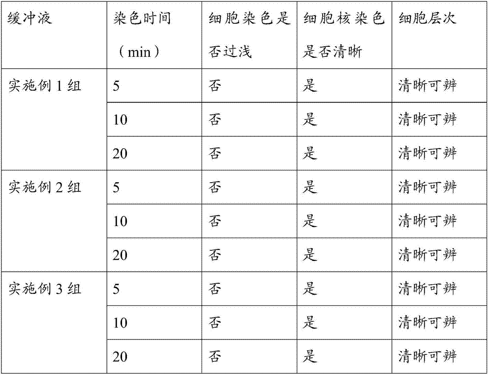
Embodiment 1
[0025] A blood cell staining method, comprising the following steps:
[0026] S1, preparation of staining solution
[0027] The staining solution was mixed with Wright's pigment, methanol and glycerin and then placed at room temperature for 3 days for later use. During the placement, it was shaken to promote the dissolution of Wright's pigment, wherein the ratio of Wright's pigment:methanol:glycerin was 1g:60ml:1ml;
[0028] S2, preparation of staining buffer
[0029] Each liter of staining buffer is prepared by mixing the following components: 0.3 g of potassium dihydrogen phosphate, 0.2 g of disodium hydrogen phosphate, 5 ml of glycerin, 5 μL of hydrogen peroxide with a mass fraction of 30%, distilled water to 1 L, and set aside;
[0030] S3, dyed
[0031] Dilute the staining solution of S1 with the staining buffer of S2, let it stand for 5 minutes after dilution, and mix well to obtain the diluted staining solution, and directly drop the diluted staining solution on the p...
Embodiment 2
[0037] A blood cell staining method, comprising the following steps:
[0038] S1, preparation of staining solution
[0039] The staining solution was mixed with Wright's pigment, methanol and glycerin and then placed at room temperature for 3 days for subsequent use. During the placement period, it was shaken to promote the dissolution of Wright's pigment, wherein the ratio of Wright's pigment: methanol: glycerol was 1g: 60ml: 2ml;
[0040] S2, preparation of staining buffer
[0041] Each liter of staining buffer is prepared by mixing the following components: 0.3 g of potassium dihydrogen phosphate, 0.2 g of disodium hydrogen phosphate, 10 ml of glycerin, 10 μL of hydrogen peroxide with a mass fraction of 30%, distilled water to 1 L, and set aside;
[0042] S3, dyed
[0043] Dilute the staining solution of S1 with the staining buffer of S2, let it stand for 5 minutes after dilution, and mix well to obtain the diluted staining solution, and directly drop the diluted staining...
Embodiment 3
[0049] A blood cell staining method, comprising the following steps:
[0050] S1, preparation of staining solution
[0051] The staining solution is mixed with Wright's pigment, methanol and glycerin and then placed at room temperature for 3 days for later use. During the placement period, it is shaken to promote the dissolution of Wright's pigment, wherein the ratio of Wright's pigment: methanol: glycerin is 1g: 60ml: 1.5ml ;
[0052] S2, preparation of staining buffer
[0053] Each liter of staining buffer is prepared by mixing the following components: 0.3 g of potassium dihydrogen phosphate, 0.2 g of disodium hydrogen phosphate, 7.5 ml of glycerin, 7.5 μL of hydrogen peroxide with a mass fraction of 30%, distilled water to 1 L, and set aside;
[0054] S3, dyed
[0055]Use the staining buffer of S2 to dilute the staining solution of S1, dilute and mix well and let it stand for 8 minutes to obtain the diluted staining solution. Add the diluted staining solution directly o...
PUM
| Property | Measurement | Unit |
|---|---|---|
| Diameter | aaaaa | aaaaa |
Abstract
Description
Claims
Application Information
 - R&D Engineer
- R&D Manager
- IP Professional
- Industry Leading Data Capabilities
- Powerful AI technology
- Patent DNA Extraction
Browse by: Latest US Patents, China's latest patents, Technical Efficacy Thesaurus, Application Domain, Technology Topic, Popular Technical Reports.
© 2024 PatSnap. All rights reserved.Legal|Privacy policy|Modern Slavery Act Transparency Statement|Sitemap|About US| Contact US: help@patsnap.com


