Garma photon detection imaging device and method

A photon and positron technology, applied in the field of Gama photon detection devices, can solve the problems of the movement of the treatment target, the high ratio of non-real time coincidence events, and the low detection efficiency, so as to save costs and improve the signal-to-noise ratio of images. Effect

Active Publication Date: 2020-03-10
CHUNG GUNG MEDICAL FOUNDATION LINKOU BRANCH
View PDF9 Cites 0 Cited by
  • Summary
  • Abstract
  • Description
  • Claims
  • Application Information

AI Technical Summary

Problems solved by technology

[0006] (2) There is a difference between the actual treatment position and the treatment plan: mainly due to the movement of the treatment target caused by the patient's positioning error, breathing or heartbeat, etc.;
[0008] (4) Errors caused by facilities that generate proton or heavy ion beams, such as errors in energy degraders, beam delivery related devices, etc.
In short, direct application of the existing DSSD-type and CZT-type probes designed for astronomy, homeland and nuclear energy safety requirements to the application of prompt gamma photon imaging in proton therapy has low detection efficiency, poor time resolution and false time-signal coincidence events The ratio is too high, which will affect the signal-to-noise ratio of the system, the image clarity and the accuracy of proton range Bragg peak estimation. Therefore, it is necessary to measure the Compton scattering imaging detection probe for the demand of proton therapy.
[0016] In addition, prior art such as U.S. Patent Publication No. US6,484,051 discloses a device capable of simultaneously detecting positron-mutually destructive Gamma photons and prompt Gamma photons produced by isotopes. Due to the need for simultaneous measurement, therefore At least three detectors are required to work at the same time
In addition, the signal of the prompt gamma photons is tens of times that of the positron mutual destructive gamma photons. If measured simultaneously, the detection counting rate (counting rate) will be lowered by the low yield rate (yield rate) of the positron mutual destructive gamma photons. Quantity dominates, which affects the signal-to-noise ratio of the detected signal, thereby reducing the quality of image reconstruction

Method used


Image

  • Garma photon detection imaging device and method
  • Garma photon detection imaging device and method
  • Garma photon detection imaging device and method

Examples


Embodiment Construction

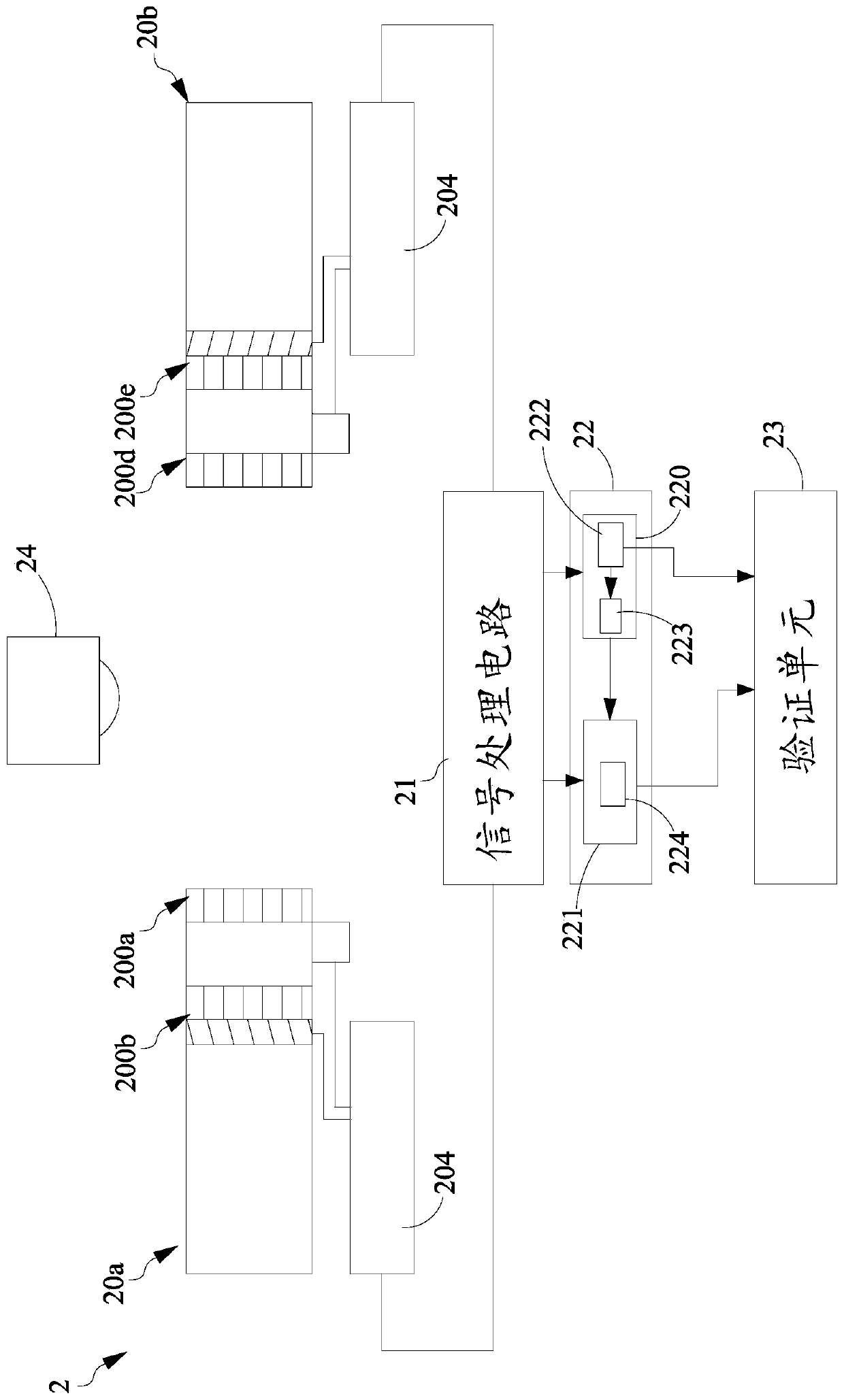
[0039] see Figure 2A As shown, this figure is a schematic diagram of the structure of an embodiment of the gamma photon detection device provided by the present invention. In this embodiment, the gamma photon detection device 2 includes a plurality of detection probes 20 a and 20 b , a signal processing circuit 21 , a reconstruction unit 22 and a verification unit 23 . Each detection probe 20a and 20b is a probe with a nanosecond response level, and its detection surfaces correspond to each other and are parallel to each other. Each detection probe 20a and 20b has multi-layer scintillation crystal detectors 200a-200b and 200d-200e, arranged along the axial direction of the corresponding detection probe, adjacent scintillation crystal detectors 200a-200b and 200d-200e has a distance. In one embodiment, each of the detection probes 20 a and 20 b is used to capture gamma photons generated by a charged particle beam along a traveling direction to generate corresponding electric...


PUM


Abstract

The invention provides an apparatus and a method for imaging gamma ray. The gamma ray detecting apparatus comprises plural detector heads, and a signal processing circuit, the plural detector heads respectively comprising plural scintillation detectors. The detector heads are utilized to detect gamma rays generated from an object. The gamma rays first work on the first layer of scintillation detector and then on the second layer of scintillation detector. Then, the first electrical signals acquired by each detector head during the first period are obtained and the first coincidence calculationon the first electrical signals corresponding to each scintillation detector is operated, thereby obtaining plural the first coincident data corresponding to each detector head. Finally, the second electrical signals of the first layer of scintillation detector in each detector are obtained during the second period, and the second coincidence calculation on the second electrical signals, therebyobtaining plural pairs of the second period coincident data. The first and second coincident signals are utilized to reconstruct activity distribution.

Description

technical field [0001] The present invention relates to a gamma photon detection device and method, in particular to a gamma photon detection imaging for detecting prompt gamma ray and positron annihilation gamma ray Devices and methods. Background technique [0002] Such as figure 1 As shown, radiotherapy (Radiotherapy, RT) is to irradiate the lesion with high-energy photons (such as X-rays) or charged particles, in order to achieve the purpose of "killing malignant tumors" or "inhibiting the proliferation of malignant tumors". treatment. Among them, the curve 90 represents the relationship between X-ray dose and tissue depth. Due to the physical characteristics of the interaction between X-ray rays and substances, as the ray enters the depth of the tissue, the relative dose also gradually decays with the depth. Therefore, the path of a single X-ray beam entering the body More doses are released on the normal tissues before and after the tumor, causing the X-rays to affe...

Claims


Application Information

Patent Timeline
Patent Type & Authority Patents(China)
IPC IPC(8): G01T1/202
Inventor 詹美龄洪志宏
Owner CHUNG GUNG MEDICAL FOUNDATION LINKOU BRANCH
Why Eureka
Try Eureka