An activatable tumor apoptosis pet imaging agent and its preparation method and application

A PET imaging agent and apoptosis technology, which is applied in the field of tumor apoptosis positron emission tomography imaging agent and its preparation, can solve the problems of poor imaging sensitivity, short retention time, and low labeling efficiency, and achieve imaging sensitivity Strong, simple preparation method, short labeling time

Active Publication Date: 2020-09-08
JIANGSU INST OF NUCLEAR MEDICINE
View PDF5 Cites 0 Cited by
  • Summary
  • Abstract
  • Description
  • Claims
  • Application Information

AI Technical Summary

Problems solved by technology

However, the aforementioned apoptosis probes [ 18 The labeling time of F]-CP18 is as long as 92 minutes. The labeling time is too long, the labeling efficiency is low, the retention time in the cells is short, the target-to-clip ratio is low, and the imaging sensitivity is poor. It is difficult to meet the current high-precision clinical requirements for early evaluation of tumor efficacy.

Method used


Image

  • An activatable tumor apoptosis pet imaging agent and its preparation method and application
  • An activatable tumor apoptosis pet imaging agent and its preparation method and application
  • An activatable tumor apoptosis pet imaging agent and its preparation method and application

Examples


Embodiment 1

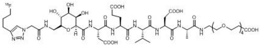
[0062] The DEVD described in this example 18 F marker has a structure shown in the following formula (1):

[0063]

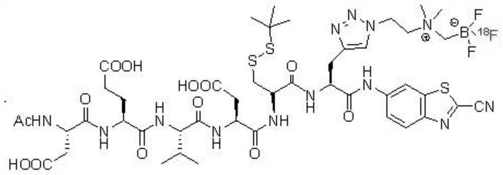
[0064] The DEVD's 18 The synthetic route of the F marker is:

[0065]

[0066] The DEVD's 18 The synthesis steps of F markers include:

[0067] (1) Add 150 mg (0.325 mmol) of the compound shown in formula (A) and 246 mg (0.380 mmol) of the polypeptide DEVD shown in formula (B) into a 25 mL round bottom flask, add 3 mL of dry tetrahydrofuran (THF) to dissolve, Then add O-benzotriazole-tetramethyluronium hexafluorophosphate (HBTU) 142mg (0.375mmol) and N,N-diisopropylethylamine (DIPEA) 142μL (0.815mmol), under nitrogen protection After reacting at room temperature for 2 hours, the above reaction solution was subjected to TLC detection, developed with dichloromethane:methanol=20:1 (v / v), detected by 254nm ultraviolet light, Rf=0.6, and spin-dried the solvent after judging that the reaction was completed, and obtained the crude product Separation and puri...

Embodiment 2

[0086] The synthesis steps of the 18F marker of the molecular probe DEVD described in this example include:

[0087] (1) Add 150 mg (0.325 mmol) of the compound represented by formula (A) and 243.75 mg (0.325 mmol) of the polypeptide DEVD represented by formula (B) into a 25 mL round bottom flask, add 3 mL of dry tetrahydrofuran (THF) to dissolve , then add O-benzotriazole-tetramethyluronium hexafluorophosphate (HBTU) 147.68mg (0.39mmol) and N, N-diisopropylethylamine (DIPEA) 124.5μL (0.715mmol), nitrogen After reacting at room temperature for 1 h under protection, the above reaction solution was subjected to TLC detection, developed with dichloromethane:methanol=20:1 (v / v), detected by 254nm ultraviolet light, Rf=0.6, and spin-dried the solvent after judging that the reaction was completed, and the The obtained crude product was separated and purified by silica gel column chromatography, using octadecylsilane bonded silica gel as filler, chromatographic column (5 μm, 250 × 4....

Embodiment 3

[0093] The DEVD described in this example 18 The synthesis steps of F markers include:

[0094] (1) Add 150 mg (0.325 mmol) of the compound represented by formula (A) and 252.5 mg (0.390 mmol) of the polypeptide DEVD represented by formula (B) into a 25 mL round bottom flask, add 3 mL of dry tetrahydrofuran (THF) to dissolve , then add O-benzotriazole-tetramethyluronium hexafluorophosphate (HBTU) 123mg (0.325mmol) and N,N-diisopropylethylamine (DIPEA) 152.9μL (0.8775mmol), nitrogen protection After reacting at room temperature for 3 hours, the above reaction solution was subjected to TLC detection, developed with dichloromethane:methanol=20:1 (v / v), detected by 254nm ultraviolet light, Rf=0.5, it was judged that the reaction was complete, and then the solvent was spin-dried, and the The resulting crude product was separated and purified by silica gel column chromatography, using octadecylsilane bonded silica gel as a filler, a chromatographic column (5 μm, 250 × 4.6 mm, Pheno...


PUM

PropertyMeasurementUnit
purityaaaaaaaaaa

Abstract

The invention belongs to the field of radioactive medicine labeling, and in particular to an activatable tumor apoptosis PET (positron emission computed tomography) developer, as well as a preparationmethod and application thereof. The activatable tumor apoptosis PET developer has a structure as shown in a formula (I), the labeling process only needs 30 minutes, the labeling time is short, the radiochemical yield of the labeling product can reach 54 percent, the specific activity is 1.2 Ci/umol, the radiochemical purity is more than 98 percent, the labeling efficiency is high, the stability is high and the preparation steps are simple. The labeling product has high in-vitro stability, has high targeting property on apoptotic cells, can serves as a PET developer to detect tumor apoptosis,and also can be applied to early diagnosis of related diseases caused by abnormal cell apoptosis, detection progress, early curative effect evaluation after treatment or development of new treatment schemes and personalized diagnosis and treatment schemes.

Description

technical field [0001] The invention belongs to the field of radiopharmaceutical labeling, and in particular relates to an activatable tumor apoptosis positron emission tomography (Positron Emission Computed Tomography, PET for short) agent and its preparation method and application. Background technique [0002] Apoptosis refers to the process of active cell death controlled by genes in order to maintain a stable internal environment. Apoptosis is different from cell necrosis. Apoptosis is not a passive process, but an active process. It involves the activation, expression, and regulation of a series of genes. It is not a phenomenon of autologous injury under pathological conditions. , but a death process that is actively fought for to better adapt to the living environment. The morphological changes of apoptosis are mainly manifested as the condensation of chromatin in the nucleus, the condensation of cytoplasm in the cytoplasm, and the appearance of biological macromolec...

Claims


Application Information

Patent Timeline
Patent Type & Authority Patents(China)
IPC IPC(8): C07K7/06C07K1/13A61K51/08A61K101/02
Inventor 邱玲林建国李珂彭莹吕高超刘清竹谢敏浩
Owner JIANGSU INST OF NUCLEAR MEDICINE
Why Eureka
Try Eureka