Infectious bovine rhinotracheitis virus gD protein antigen epitope polypeptide, and inhibitor, monoclonal antibody and application of polypeptide

A technology of rhinotracheitis virus and protein antigen, applied in the fields of molecular biology and medicine, can solve the problems of economic loss, secondary bacterial infection, disease control and other problems in the cattle industry

Inactive Publication Date: 2018-01-16
HAINAN UNIVERSITY
View PDF3 Cites 5 Cited by
  • Summary
  • Abstract
  • Description
  • Claims
  • Application Information

AI Technical Summary

Problems solved by technology

The disease has a wide host range, which restricts animal trade and brings huge economic losses to the development of the cattle industry
Although the fatality rate of this disease is not high, seco

Method used


Image

  • Infectious bovine rhinotracheitis virus gD protein antigen epitope polypeptide, and inhibitor, monoclonal antibody and application of polypeptide
  • Infectious bovine rhinotracheitis virus gD protein antigen epitope polypeptide, and inhibitor, monoclonal antibody and application of polypeptide
  • Infectious bovine rhinotracheitis virus gD protein antigen epitope polypeptide, and inhibitor, monoclonal antibody and application of polypeptide

Examples


Embodiment 1

[0032] The preparation of embodiment 1 monoclonal antibody

[0033] 1. Immunization of mice

[0034] Immunization of mice Three 4-6-week-old female BALB / c mice were immunized with prokaryotically expressed recombinant gD-pET-28a protein purified by cutting gel, and immunized three times in total, with two weeks between each immunization, and the immunization dose was 60 μg / Only, for the first time, an equal amount of Freund's complete adjuvant was used to emulsify with protein, and for the second and third times, an equal amount of Freund's incomplete adjuvant was used for emulsification, and the immunization route was subcutaneous immunization. Immunization was boosted by intraperitoneal injection of 30 μg purified recombinant protein (without any adjuvant) before fusion.

[0035] 2. Cell Fusion

[0036] The feeder layer cells were prepared 1 day before fusion, and BALB / c mouse peritoneal macrophages were plated in 96-well cell culture plates according to conventional meth...

Embodiment 2

[0039] Identification of embodiment 2 monoclonal antibody

[0040] 1. Subclass identification of monoclonal antibodies

[0041] The subclass of the anti-gD protein monoclonal antibody 2B6 was identified as IgG2a / κ by the antibody subclass identification kit.

[0042] 2. Western blot test

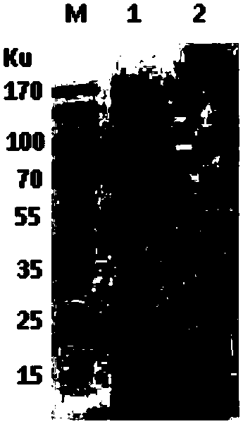
[0043] The reactivity of monoclonal antibody and recombinant protein was verified by Western blot. The purified recombinant protein was transferred to PVDF membrane after SDS-PAGE, the positive hybridoma cell line 2B6 culture supernatant was used as the primary antibody, HRP-labeled goat anti-mouse IgG was used as the secondary antibody, and DAB was used for color development ( figure 1 ).

[0044] The reactivity of monoclonal antibody with IBRV was verified by Western blot. The virus purified by sucrose gradient centrifugation was transferred to PVDF membrane, the positive hybridoma cell line 2B6 culture supernatant was used as the primary antibody, HRP-labeled goat anti-mouse IgG was u...

Embodiment 3

[0048] Example 3 Screening of antigenic epitopes

[0049] 1. Preliminary identification of antigenic epitopes

[0050] Referring to the amino acid sequence of BHV-1gD, the experiment designed 3 pairs of primers to divide the gD protein into 3 segments, each segment has partial amino acid overlap, and the upstream of each pair of primers was inserted into the BamH Ⅰ site, and the downstream of each pair was inserted into the EcoRI site. After amplification and purification by PCR, enzyme digestion and connection with pGEX-6P-1 vector, (PCR system, enzyme digestion system and connection system are shown in Table 2-5) the recombinant protein containing GST tag was expressed, and each fusion short was determined by Western blot. Reactivity of peptides with MAb 2B6. After thrice truncated expression ( Figure 4 ), the size of the epitope was preliminarily determined to be 15 amino acids, namely 323 GEPKPGPSDADRPE 337 .

[0051] Table 1 Design of primers for preliminary epitope...


PUM


Abstract

The invention belongs to the field of molecular biology and medicine, and specifically discloses an infectious bovine rhinotracheitis virus gD protein antigen epitope polypeptide and an application ofthe polypeptide in preparation of a reagent or a medicament for detecting or treating infectious bovine rhinotracheitis. The invention provides a monoclonal antibody for resisting the infectious bovine rhinotracheitis virus gD protein, and simultaneously the antigen epitope of the infectious bovine rhinotracheitis virus gD protein is screened out as <323>GEPKPGPSPDADRPE<337> (the shortest epitopesequence is 7 amino acid peptide fragments: <323>GEPKPGP<329>). The recombinant protein based on the antigen epitope can specifically be used to detect infectious bovine rhinotracheitis serum; in addition, a small-molecule inhibition drug designed based on the antigen epitope can block virus infection; and meanwhile, the multi-copy repeated epitope vaccine constructed on the basis of the antigenepitope can induce a high-titer gD protein antibody under the assistance of an appropriate adjuvant, and has a relatively high neutralizing antibody titer. The invention lays a foundation for establishing a detection method and researching and developing vaccines for the infectious bovine rhinotracheitis.

Description

technical field [0001] The invention belongs to the fields of molecular biology and medicine, and in particular relates to a bovine infectious rhinotracheitis virus gD protein antigen epitope polypeptide and its use in preparing reagents or medicines for detecting or treating bovine infectious rhinotracheitis. Background technique [0002] Infectious Bovine Rhinotracheitis (IBR) is an important respiratory disease of cattle caused by Infectious Bovine Rhinotracheitis virus (IBRV) that can lead to weight loss, miscarriage, milk production Drops, bilateral conjunctivitis, and extreme restlessness. Clinically, it can be divided into respiratory tract type, reproductive tract infection type, encephalitis type, ophthalmia type and abortion type. The main routes of disease transmission are direct contact with pathogens and latent infection. The disease has a wide host range, which restricts animal trade and brings huge economic losses to the development of the cattle industry. ...

Claims


Application Information

Patent Timeline
IPC IPC(8): C07K14/06C07K16/08C12N5/20G01N33/68G01N33/569A61K39/265A61K39/42A61K45/00A61P31/22A61P11/00
Inventor 倪宏波
Owner HAINAN UNIVERSITY
Why Eureka
Try Eureka