
Intravascular endoscopic ultrasound-OCT probe system

An ultrasound and vascular technology, applied in the field of medical endoscopy, can solve the problems of electromagnetic interference, limited penetration depth, not on the same section, etc., and achieve the effect of high safety and high sensitivity

Inactive Publication Date: 2018-02-23
TIANJIN UNIV
View PDF6 Cites 8 Cited by
  • Summary
  • Abstract
  • Description
  • Claims
  • Application Information

AI Technical Summary

Problems solved by technology

However, due to the limited penetration depth of intravascular OCT (field of view radius 3-4mm, penetration depth 1-2mm), its ability to identify calcified and lipid plaques is not strong
(The full picture of vulnerable plaques with large lipid cores cannot be fully described
[0004] Patent Application No. 201410333307.3 The imaging probe with a combined ultrasonic and optical imaging device introduces an ultrasonic-OCT imaging probe driven by an external proximal drive device. The external drive has certain electromagnetic interference, and it is not easy to control the insertion of the distal end into the blood vessel. The swing amplitude of the inner imaging part is less secure
[0005] Patent authorization announcement number CN1322839C invented a medical endoscope micro-ultrasound-OCT probe, the ultrasonic transducer and the OCT imaging device are located at both ends of the ultrasonic motor, and the images formed are not on the same cross-section. In addition, the invention The image processing and display part connected to the imaging probe is not described

Method used


Image

  • Intravascular endoscopic ultrasound-OCT probe system
  • Intravascular endoscopic ultrasound-OCT probe system

Examples


Embodiment Construction

[0023] Embodiments of the present invention are described in detail below in conjunction with the accompanying drawings:

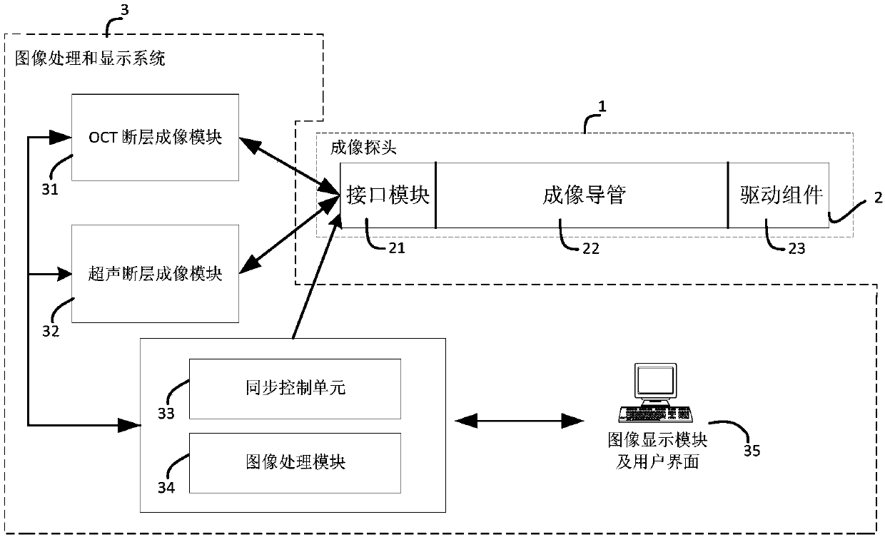
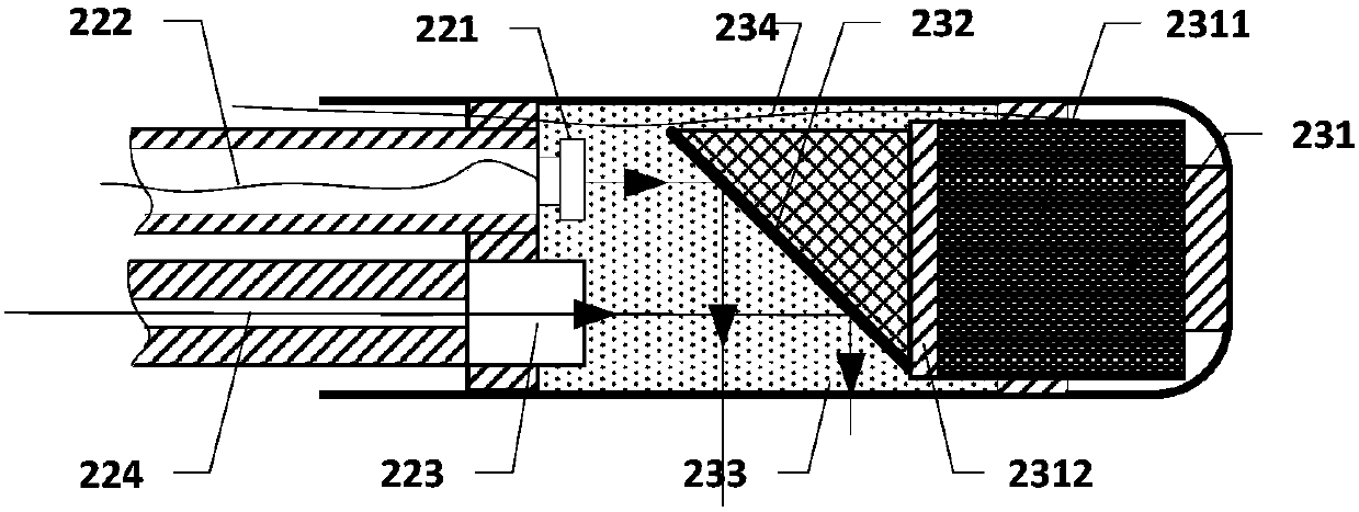
[0024] like figure 1 and figure 2 As shown, the present invention protects an endovascular ultrasound-OCT probe system, which is used for inserting an imaging probe 1 into an arterial vessel for imaging. The hollow catheter 2 has a distal portion, a proximal portion and a middle portion, a drive assembly 23 is disposed at the distal portion within the elongated hollow catheter, and the proximal side of the drive assembly 23 fixes an imaging catheter 22 passing through the elongated hollow catheter The middle part of 2 and the interface module 21 encapsulated in the proximal part of the elongated hollow conduit 2 are connected to the image processing and display system 3;

[0025] The drive assembly 23 includes an ultrasonic motor 231, an acousto-optic mirror 232, the ultrasonic motor 231 includes an ultrasonic motor stator 2311 and an ultrasonic motor r...


PUM


Abstract

The invention discloses an intravascular endoscopic ultrasound-OCT probe system, which is applied to imaging in an artery blood vessel. The probe system comprises an imaging probe which is constitutedby a slender hollow catheter, wherein a driving component, an imaging catheter and an interface module are sequentially arranged on a far-end part, a middle part and a near-end part of the interior of the slender hollow catheter, and the interface module is connected to an image processing and display system; the image processing and display system comprises an OCT tomography module, an ultrasonic tomographic image imaging module, a synchronous control unit, an image processing module, an image display module and a user interface; the driving component comprises an ultrasonic motor and an acousto-optical reflector; the ultrasonic motor is composed of an ultrasonic motor stator and an ultrasonic motor rotor; the imaging catheter comprises an OCT probe catheter part and an ultrasonic imaging catheter part; and the OCT probe catheter part is composed of a single mode fiber and a grin lens. According to the probe system provided by the invention, a near-end driving device is arranged at an ultrasound inner side and is free from electromagnetic interference; and meanwhile, the swinging amplitude of an imaging part that the far end is inserted into a blood vessel can be easily controlled, so that higher safety is guaranteed.

Description

technical field [0001] The invention relates to the field of medical endoscopy, in particular to a vascular endoscope ultrasound-OCT probe system. Background technique [0002] The major cause of most acute coronary heart disease and intravascular thrombosis is the rupture of vulnerable plaques (thin fibrous cap lipid plaques, TCFAs) attached to the vessel wall. Long-term studies on patients with coronary heart disease have shown that TCFA has the characteristics of thin fibrous cap, large lipid core, rich in macrophages, glycoprotein matrix, inflammatory response, and calcified nodular plaques, while the thickness of fibrous cap of TCFA is closely related to lipid. The size of the mass is the main parameter to characterize its hazard. [0003] At present, the main method for detecting coronary heart disease is coronary angiography, but this technology can only image the intravascular blood flow. The current imaging methods for morphological examination of vulnerable plaqu...

Claims


Application Information

Patent Timeline
IPC IPC(8): A61B5/00A61B8/12A61B8/08
CPCA61B5/0066A61B5/0084A61B5/6876A61B5/72A61B8/0891A61B8/12A61B8/5207A61B8/5261
Inventor 陈晓冬陈晓帅汪毅李嘉科郁道银
Owner TIANJIN UNIV
Learn More