Preparation method of immunohistochemical detecting section

A technique of immunohistochemistry and tissue slicing, which is applied in the field of preparation of immunohistochemical detection slices, which can solve the problems of difficulty in satisfying diversity detection and the complexity of slice production, and achieve the effects of avoiding reagent evaporation, reducing reagent consumption, and easy humidity

Active Publication Date: 2018-05-15
潘永红
View PDF5 Cites 0 Cited by
  • Summary
  • Abstract
  • Description
  • Claims
  • Application Information

AI Technical Summary

Problems solved by technology

[0003] Due to the small number of clinical tissue samples in actual testing, the complexity of making slices, and the need to perform multiple antigens or antibodies or other related tests on the same tissue sample to be tested, multiple detection slices are required. Therefore, The traditional detection slice preparation method is difficult to meet the needs of subsequent diversity detection

Method used


Image

  • Preparation method of immunohistochemical detecting section
  • Preparation method of immunohistochemical detecting section
  • Preparation method of immunohistochemical detecting section

Examples


Embodiment 1

[0030] This embodiment provides a method for preparing immunohistochemical detection slices, comprising the following steps:

[0031] Step 1: Obtain a paraffin section of the breast tissue sample to be tested, and spread the paraffin section on a polyimide plastic film (commercially available, with a tolerance temperature of 400°C and an embrittlement temperature of minus 200°C), and paraffin section Tightly adhere to the plastic film to form a paraffin section attached to the plastic film, and bake the slice at 60°C for 30 minutes after rolling; the thickness of the polyimide plastic film used in this step is 30 μm; the rolling method described in this step is to use The rubber roller rolls back and forth on the paraffin section of breast cancer;

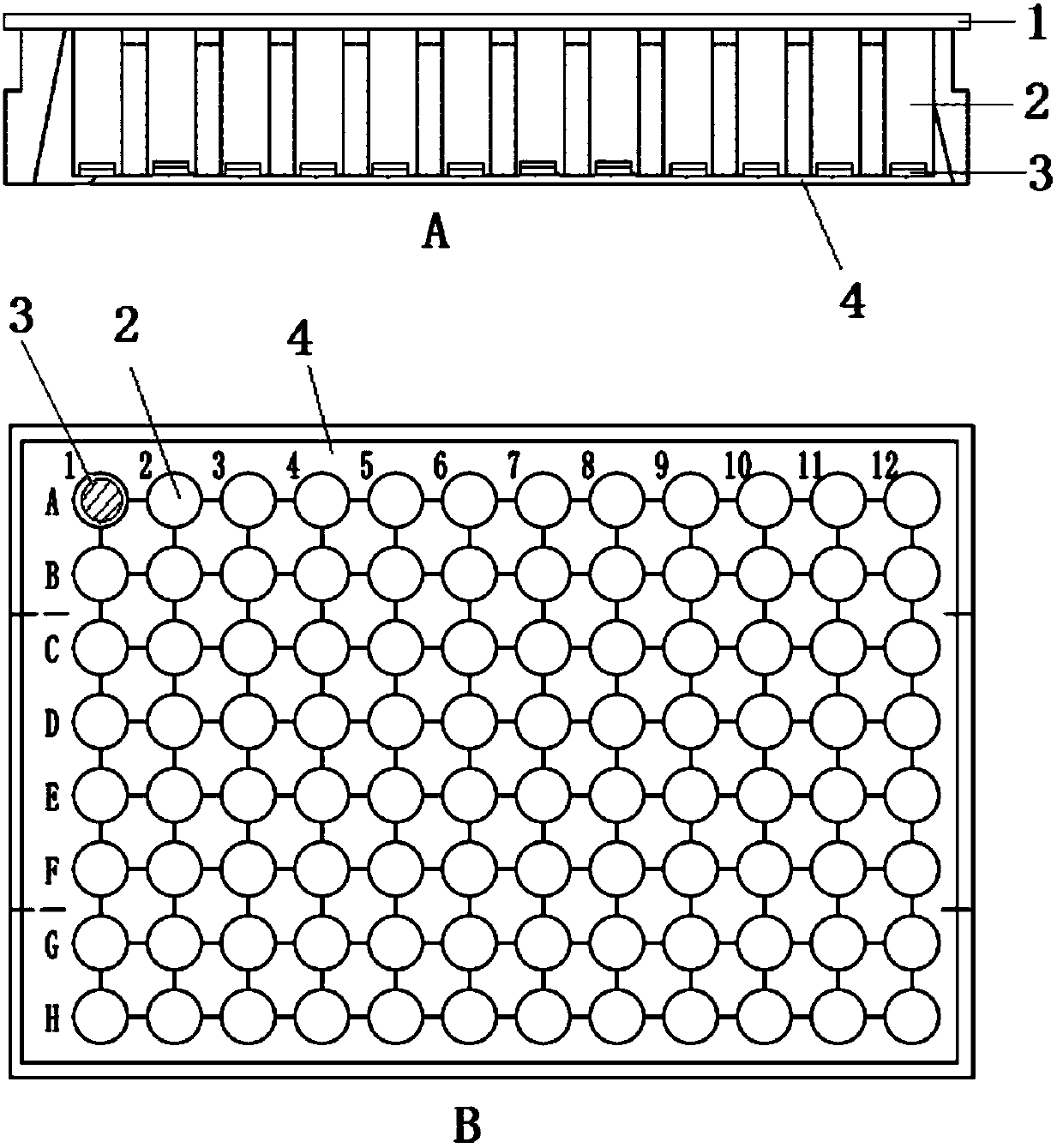
[0032] Step 2, punching the obtained paraffin section attached with a plastic film to obtain 18 (6 treatments in total, each treatment repeated 3 times) circular paraffin section attached with a plastic film whose diameter is 3mm; ...

Embodiment 2

[0049] This embodiment is a modification of Embodiment 1. Compared with Embodiment 1, the only changes are:

[0050] In step 1, the plastic film is selected polynaphthyl ester plastic film (commercially available, the tolerance temperature is 265 ℃, and the brittle temperature is lower than minus 196 ℃), the baking sheet temperature is 65 ℃, and the thickness of the polyester plastic film is 250 μm; this step The flattening method is to use a glass rod to roll back and forth on the paraffin section of breast cancer;

[0051] In step 2, the obtained paraffin section attached with a plastic film is punched to obtain a circular paraffin section attached with a plastic film with a diameter of 20 mm.

[0052] In this example, four six-well plates were used for immunofluorescent staining.

[0053] In the process of preparing 100 tissue sections according to the tissue section preparation method provided in this example, only 5 tissue sections had the plastic film fall off during th...

Embodiment 3

[0055] This embodiment is an improved example of embodiment 1. Compared with embodiment 1, the improvements only lie in:

[0056] In step 1, the plastic film is also subjected to the following pretreatment: first, the surface of the plastic film is coated with a mixture of egg white and anionic polyelectrolyte aqueous solution (the mass concentration of the aqueous solution is 0.5%) at a volume ratio of 5:2. drying, and then coating a cationic polyelectrolyte solution (the mass concentration of the aqueous solution is 0.5%) on the surface of the plastic film pre-laid paraffin section and drying, wherein the anionic polyelectrolyte is polyacrylic acid, and the cationic polyelectrolyte is polyvinylamine. The drying described in this example is air drying.

[0057] In this example, the above-mentioned pretreatment of the plastic film can not only improve the firmness of the combination of the tissue section and the plastic film, but also the plastic film has good hydrophilic prop...


PUM

PropertyMeasurementUnit
Diameteraaaaaaaaaa
Diameteraaaaaaaaaa
Temperature toleranceaaaaaaaaaa

Abstract

The invention discloses a preparation method of an immunohistochemical detecting section. The preparation method comprises the following steps: obtaining a paraffin section of a tissue sample to be detected, flatly laying the paraffin section on a plastic film, making the paraffin section and the plastic film be closely bonded to form the paraffin section attached with the plastic film and bakingthe paraffin section; punching the paraffin section attached with the plastic film after baking, and obtaining at least one round paraffin section attached with the plastic film and with the diameterbeing 1-30mm; transferring the round paraffin section attached with the plastic film into an enzyme-labeled board, setting one section for each hole, dewaxing and hydrating to obtain a round hydrophilic tissue section attached with the plastic film; dyeing the round hydrophilic tissue section attached with the plastic film to obtain the immunohistochemical detecting section. The preparation methoddisclosed by the invention has the beneficial effects that the plastic film and the paraffin section are creatively and closely bonded together by an inventor, then the plurality of paraffin sectionsare obtained by punching, the preparation of multiple sections by adoption of one paraffin section of tissue to be detected is finally realized, and the need of multiple detection is met simultaneously.

Description

technical field [0001] The invention relates to the field of biotechnology, in particular to a method for preparing immunohistochemical detection slices. Background technique [0002] Pathological examination refers to a kind of pathological examination, which is a pathological method for examining pathological changes in organs, tissues or cells of the body. At present, biopsy has been widely used in clinical work and scientific research, and the preparation of microscopic biopsy plays a key role in the diagnosis of the results. The preparation of traditional microscopy sections is mainly to take pathological tissues with a size of 2.0cm×2.0cm×0.3cm and embed them in paraffin, then slice them to obtain paraffin sections, and then attach the whole piece of paraffin sections to the carrier. Staining is carried out on slides, and each whole paraffin section can only be stained once to prepare a microscopic examination section, and finally obtain one result. [0003] Due to t...

Claims


Application Information

Patent Timeline
IPC IPC(8): G01N33/531
CPCG01N33/531
Inventor 丁晓昆黄若磐丁勃
Owner 潘永红
Why Eureka
Try Eureka