Tubular material with X ray developing function, and preparation method thereof

An X-ray and tube technology, used in the medical field, can solve the problems of material performance decline and poor surface smoothness of medical catheters, and achieve excellent performance, excellent internal and external surface quality and smooth effects.

Inactive Publication Date: 2018-07-13
SHENZHEN POLYTECHNIC +1
View PDF14 Cites 8 Cited by
  • Summary
  • Abstract
  • Description
  • Claims
  • Application Information

AI Technical Summary

Problems solved by technology

[0005] The present invention aims to provide a tube with X-ray imaging function and its preparation method, which can solve the technical problems of poor surface smoothness and material performance degradation of existing medical catheters with X-ray imaging function

Method used


Image

  • Tubular material with X ray developing function, and preparation method thereof
  • Tubular material with X ray developing function, and preparation method thereof
  • Tubular material with X ray developing function, and preparation method thereof

Examples


preparation example Construction

[0034] The embodiment of the present invention also provides a method for preparing the above-mentioned tube with X-ray imaging function, which includes the following steps:

[0035] 1. Add 34.2-60.2% of base resin, 39-65% of developer, 0.1-0.5% of antioxidant, 0.1-0.4% of dispersant, 0.1-0.3% of internal lubricant, and 0.1-0.3% of external lubricant into high-speed mixing In the machine, mix for 3-5 minutes;

[0036] 2. Add the mixed materials into a twin-screw extruder for melt blending, extrusion and granulation to obtain a medical catheter composite material with an X-ray imaging function with a developer weight content of 39-65%. Wherein, the temperature of each working section of the twin-screw extruder is set according to the melting point or melting temperature of the base resin;

[0037] 3. Use two single-screw extruders to form a composite extruder, and extrude with a co-extrusion head to obtain a pipe with a three-layer structure.

[0038] 3.1 sending the composit...

Embodiment 1

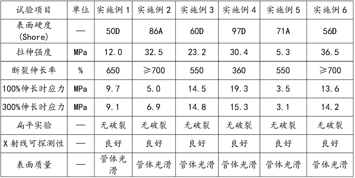
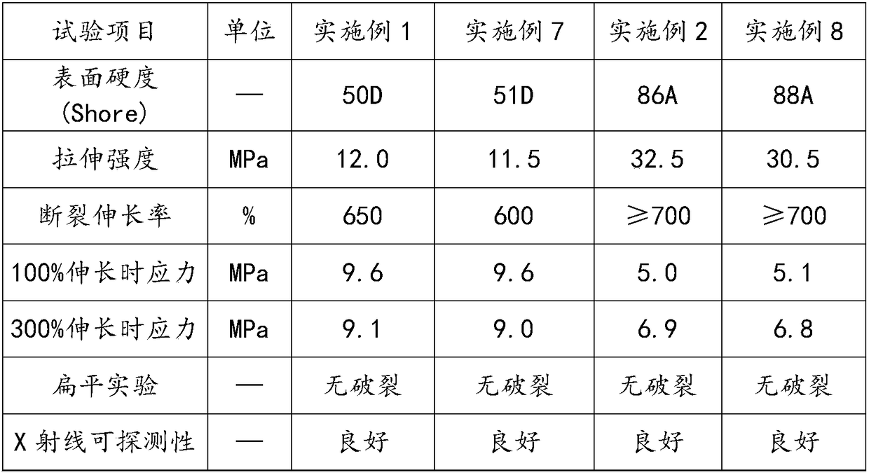
[0043] This embodiment provides a low-density polyethylene composite pipe with X-ray imaging function, and its raw material composition and preparation method are as follows:

[0044] 1. Low-density polyethylene 60.2%, bismuth trioxide 39%, antioxidant 1010 0.15%, antioxidant 1680.15%, diffusing agent EBS 0.2%, zinc stearate 0.15% and polyethylene wax 0.15%; In a high-speed mixer, dry mix for 5 minutes to obtain a mixed material.

[0045] 2. Add the mixed materials into a twin-screw extruder for melt blending, extrusion and granulation to obtain a low-density polyethylene composite material for the core layer. Among them, the temperature of each section of the twin-screw extruder starts from the feeding port and is set to 125°C, 170°C, 185°C, 190°C, 190°C, 190°C, 190°C, 190°C, 190°C, 190°C. The temperature was set to 185°C.

[0046] 3. Use two single-screw extruders to extrude the low-density polyethylene composite material for the core layer and the low-density polyethylene...

Embodiment 2

[0050] This embodiment provides a TPU composite pipe with X-ray imaging function, its raw material composition and preparation method are as follows:

[0051] 1. Polyurethane thermoplastic elastomer (TPU) 49.3% (the TPU is specifically Estane58315 provided by Lubrizol), barium sulfate 50%, antioxidant 1010 0.15%, diffusing agent EBS 0.25%, zinc stearate 0.15%, polyethylene Wax 0.15%; in a high-speed mixer, dry blend for 5 minutes to obtain a mixed material.

[0052] 2. Add the mixed materials into a twin-screw extruder for melt blending, extrusion and granulation to obtain a low-density polyethylene composite material for the core layer. Among them, the temperature of each section of the twin-screw extruder starts from the feeding port, and is set to 125°C, 170°C, 180°C, 185°C, 185°C, 185°C, 185°C, 185°C, 190°C, 190°C. The temperature was set to 185°C.

[0053] 3. Extrude the TPU composite material for the core layer and the TPU base resin with two single-screw extruders, an...


PUM

PropertyMeasurementUnit
diameteraaaaaaaaaa
thicknessaaaaaaaaaa
thicknessaaaaaaaaaa

Abstract

The invention belongs to the technical field of medicine, and especially relates to a tubular material with X ray developing function, and a preparation method thereof. The tubular material comprisesa core layer, an external skin layer, and an internal skin layer; the core layer is arranged between the external skin layer and the internal skin layer; the external skin layer and the internal skinlayer are prepared from a basic resin; the core layer comprises the basic resin and a developing agent. The external skin layer and the internal skin layer are basic resin layers, and contain no or little developing agent, so that excellent internal surface and external surface quality and smoothness are provided for the tubular material, and defects such as surface irregularity, burrs, pits, andparticles caused by direct adding of a large amount of the developing agent into the tubular material are avoided.

Description

technical field [0001] The embodiments of the present invention relate to the field of medical technology, in particular to a tube with X-ray imaging function and a preparation method thereof. Background technique [0002] With the improvement of the accuracy of medical diagnosis, interventional diagnosis and treatment are paid more attention, and medical catheter is a necessary device for interventional diagnosis and treatment, including: coronary artery dilatation catheter, angiography catheter, microcatheter, arteriovenous catheter sheath, catheter for nutrient infusion , Cardiac output measurement catheter, etc. Therefore, in medical operations, medical catheters are required to have an X-ray imaging function. [0003] In order to reduce the influence of external substances, the existing medical catheters are generally made of special materials with X-ray imaging function by adding inorganic developer into the polymer material matrix, and then molded into medical tubing...

Claims


Application Information

Patent Timeline
Patent Type & Authority Applications(China)
IPC IPC(8): C08L23/06C08L75/04C08L23/12C08L53/02C08L83/04C08K13/02C08K3/22C08K3/30C08K5/526C08K5/134C08K5/20C08K5/098F16L9/14F16L11/00B32B27/32B32B27/18B32B27/06B32B27/40B32B27/30B32B27/34B32B1/08B29C47/06B29C47/00B29C48/09B29C48/18
CPCF16L9/14F16L11/005C08L23/06C08L23/12C08L53/025C08L75/04B29C48/09B29C48/18B32B1/08B32B27/08B32B27/18B32B27/30B32B27/32B32B27/34B32B27/40C08L2207/066C08L2207/062C08K2003/3045C08L2205/035C08L2205/025C08L2203/18B32B2535/00B32B2597/00C08K13/02C08K3/22C08K5/1345C08K5/20C08K5/098C08K3/30C08L83/04C08K5/526C08L21/00
Inventor 肖望东李伯全张文凯林峰徐令军
Owner SHENZHEN POLYTECHNIC
Why Eureka
Try Eureka