Tissue engineering acellular vascular scaffold preparation method

A vascular stent and tissue engineering technology, applied in the field of tissue engineering decellularized vascular stent preparation, can solve the problems of disease transmission risk limitation and difficult implementation, and achieve the effects of reducing immune response, improving mechanical properties, and improving vascular mechanical properties.

Inactive Publication Date: 2018-08-24
NANKAI UNIV
View PDF1 Cites 11 Cited by
  • Summary
  • Abstract
  • Description
  • Claims
  • Application Information

AI Technical Summary

Problems solved by technology

[0002] Cardiovascular disease is the number one killer of human health, and the first choice of treatment for more than half of the patients is vascular transplantation, but it is difficult for some patients to implement it due to the limitation of their own blood vessel sources
Allogeneic or xenograft blood vessels have been used as a substitute for autologous blood vessels to repair diseased or missing blood vessels, but the potential risk of disease transmission, acute immune rejection and limited donor sources limit their application in transplantation

Method used


Image

  • Tissue engineering acellular vascular scaffold preparation method
  • Tissue engineering acellular vascular scaffold preparation method
  • Tissue engineering acellular vascular scaffold preparation method

Examples


Embodiment 1

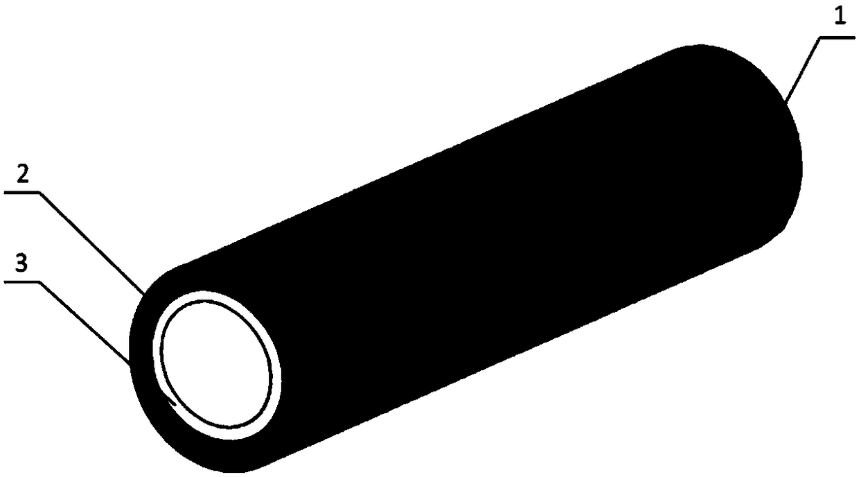
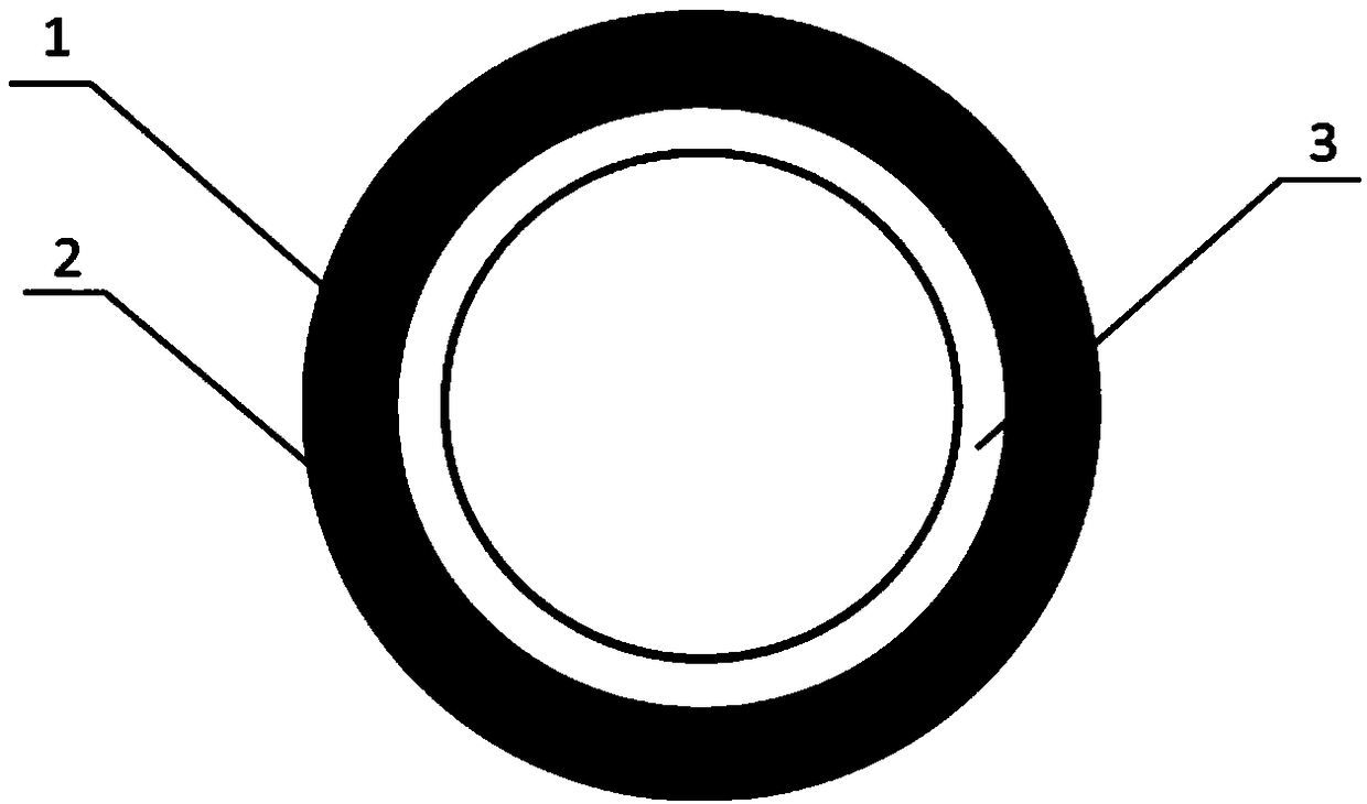
[0025] 1) Using biodegradable polymer materials as raw materials, wet spinning technology is used to prepare micron-scale or nano-scale ordered reticular vascular stents on medical silicone tubes. External helical winding, the angle between the fibers is 45°, the thickness of the mesh vascular stent layer is 600 μm, and the diameter of the vascular stent is 2mm; figure 1 It is a schematic diagram of the three-dimensional structure of decellularized blood vessels; figure 2 It is an enlarged schematic diagram of the cross-sectional structure of the decellularized vascular stent; specifically, dissolve 1 gram of PLCL in 10 mL of hexafluoroisopropanol, stir at room temperature until completely dissolved, connect a medical silicone tube with a diameter of 2 mm to a rotating motor, and inhale the PLCL solution In the syringe, the needle of the syringe is placed in an ethanol coagulation bath at a position 1 cm away from the silicone tube. Set the solution flow rate to 2ml / h, recei...

Embodiment 2

[0029] The preparation method of embodiment 2 is the same as that of embodiment 1, the only difference is that the preparation method of the spinning fiber skeleton is different: in this embodiment, the composite mandrel of melt spinning and medical silicone tube is prepared by using melt spinning equipment, and the melt spinning Wire equipment, including syringes, stainless steel single-tube needles and cylindrical receivers, in this embodiment, medical silicone tubes are sheathed with stainless steel rods to provide sufficient support to prevent the cylindrical structure from being squeezed and deformed; stainless steel syringes 1 are equipped with Spinning polymer melt; the polymer melt flows out from the all-stainless steel single-tube needle of the syringe, and rotates and translates with the cylindrical receiver, and the fine stream of polymer melt used for spinning is stretched and thinned, by adjusting the cylinder The angle of fiber arrangement can be adjusted by adjus...

Embodiment 3

[0032]Embodiment 3: embodiment 3 is similar to embodiment 2, and the difference is only that the preparation method of melt-spun fiber skeleton is different, takes by weighing 5.0g, and number average molecular weight is 80000PCL, places the sealing rust-free that fuser wraps In a steel syringe, heat at 200 °C for 1 h. Put a medical silicone tube with an outer diameter of 2mm and an inner diameter of 1mm on a stainless steel rod with a diameter of 1mm and connect it to the rotating motor. The distance between the syringe needle and the receiver is 5mm, the flow rate of the PCL melt is 0.1ml / h, and the rotation of the receiving rod The speed was set to 300r / min, the translational speed was set to 4mm / s, and the receiving device horizontally reciprocated to receive the fiber 20 times, and a composite mandrel of PCL melt-spun fiber and medical silicone tube with a fiber angle of 30° was prepared. The diameter of the spun fiber is 5 μm, and the thickness of the fiber skeleton is 4...


PUM

PropertyMeasurementUnit
diameteraaaaaaaaaa
thicknessaaaaaaaaaa
thicknessaaaaaaaaaa

Abstract

The invention belongs to the field of medical equipment, and more specifically relates to a tissue engineering acellular vascular scaffold preparation method. The method comprises the following steps:1) preparing a spinning fiber skeleton; winding the spinning fibers through a spinning technology at a receiver, and interlacing the spinning fibers up and down to form a cylindrical structure; 2) implanting the spinning fiber skeleton obtained in the step 1) under animal skin, and finally forming the host cell and extracellular matrix-coated tissue engineering vascular scaffold; and 3) removingthe receiver, and removing cells of the tissue engineering vascular scaffold. The constructed fiber skeleton is moved under the animal skin, the tissue vascular scaffold formed by migration cells dueto an immunization mechanism is constructed in the host animal body, accellular processing is carried out, the netted scaffold is kept, the blood vessel mechanical properties can be improved, an immunization reaction after transplantation can be reduced, the extracellular matrix is fully used, and good environment is provided for cell regeneration.

Description

technical field [0001] The invention belongs to the field of medical devices, and in particular relates to a method for preparing a tissue-engineered decellularized vascular stent. Background technique [0002] Cardiovascular disease is the number one killer of human health, and the first choice for more than half of patients is vascular transplantation, but it is difficult for some patients to implement it due to the limitation of their own blood vessel sources. Allogeneic or xenograft blood vessels have been used as a substitute for autologous blood vessels to repair diseased or missing blood vessels, but the potential risk of disease transmission, acute immune rejection and limited donor sources limit their application in transplantation. Contents of the invention [0003] The purpose of the present invention is to overcome the defects in the prior art and provide a preparation method of tissue engineering decellularized vascular scaffold. [0004] In order to achieve ...

Claims


Application Information

Patent Timeline
Patent Type & Authority Applications(China)
IPC IPC(8): A61L27/18A61L27/36A61L27/50A61L27/58
CPCA61L27/3633A61L27/18A61L27/3683A61L27/507A61L27/58A61L2430/40
Inventor 王恺孔德领程曲汉朱美峰张秋影傅迪宏
Owner NANKAI UNIV
Why Eureka
Try Eureka