Nanoparticle with core-shell structure, preparation method and application

A nanoparticle, core-shell structure technology, applied in the field of biomedical imaging, can solve the problem that a single modality imaging method cannot provide enough information, achieve improved water solubility and biocompatibility, simple preparation method, and less nephrotoxicity Effect

Active Publication Date: 2020-10-16
INST OF HIGH ENERGY PHYSICS CHINESE ACAD OF SCI
View PDF5 Cites 0 Cited by
  • Summary
  • Abstract
  • Description
  • Claims
  • Application Information

AI Technical Summary

Problems solved by technology

Imaging technologies have their own advantages and limitations. Single modality imaging methods usually cannot provide enough information to meet the high requirements of clinical diagnosis and diagnostic efficiency and accuracy. There is an urgent need to effectively combine various technologies

Method used


Image

  • Nanoparticle with core-shell structure, preparation method and application
  • Nanoparticle with core-shell structure, preparation method and application
  • Nanoparticle with core-shell structure, preparation method and application

Examples


Embodiment 1

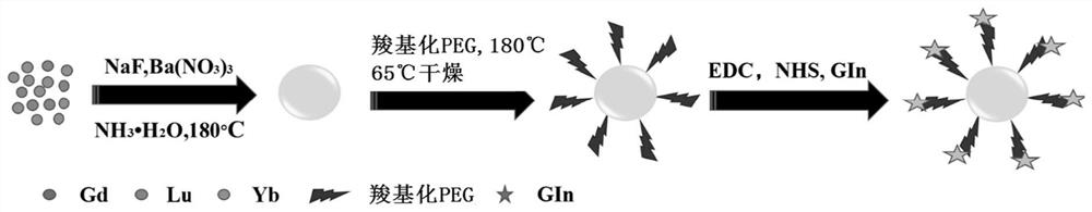
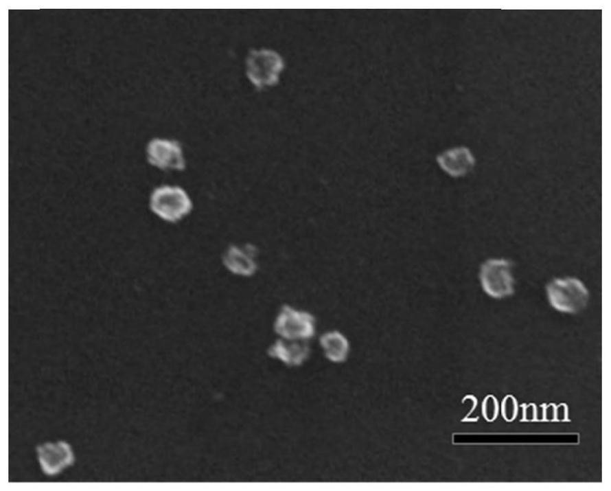
[0053] Such as figure 1 As shown, the preparation of nanoparticles with a core-shell structure includes the following steps:

[0054] (1) Ba 4 Yb 3 f 17 Preparation of Doped Gd / Lu Nanoparticles

[0055] Take a total of 1mM Yb(NO 3 ) 3 , Lu(NO 3 ) 3 , Gd(NO 3 ) 3 (molar ratio 80:12:8), inject 15mL of deionized water (DI), stir at room temperature for 10min, then add a few drops of NH 3 ·H 2 O to make the pH = about 8, after stirring for 1 h, add 5 mL of 2 mM Ba(NO 3 ) 2 Stir for another 30min, then add 15mL (20mM) NaF solution and continue stirring, mix well, put the mixed solution into the reaction vessel, heat and react at 180°C for 24h, cool to room temperature naturally, centrifuge the reaction product at 9000rpm for 10min and wash with DI Three more times to remove other residues.

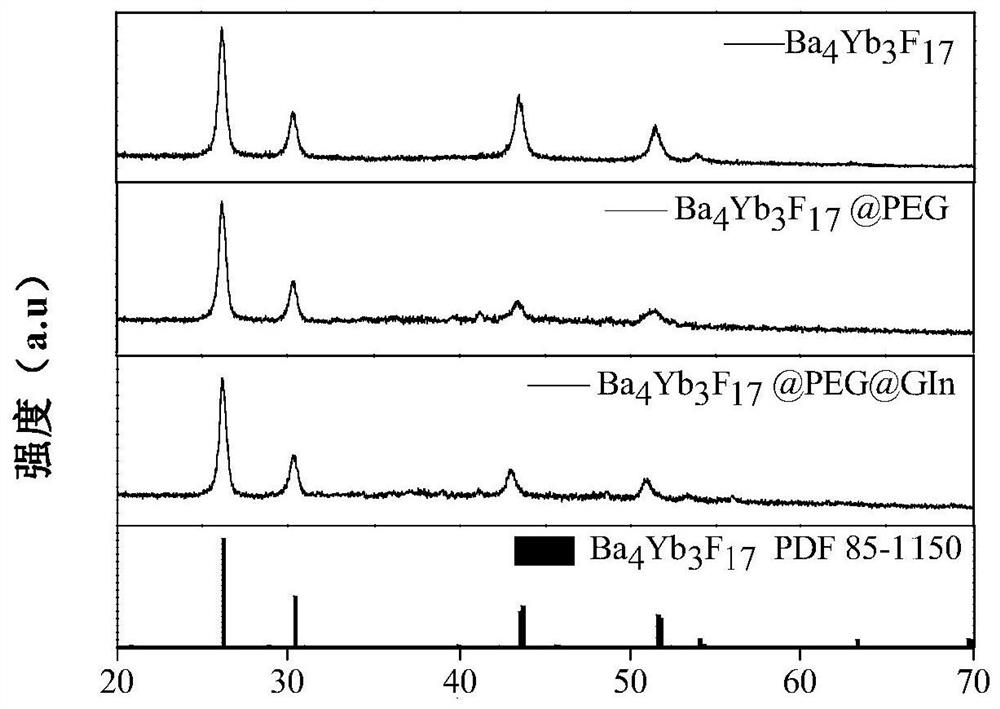
[0056] Such as image 3 As shown in the XRD diagram of the standard database, the comparison of the XRD diagram in the standard database in the bottom column shows that the dopi...

Embodiment 2

[0065] The nanomaterial Ba that embodiment 1 makes 4 Yb 3 f 17 : Gd / Lu@PEG@GIn and excess RITC were stirred at room temperature, protected from light for 4 hours, washed by centrifugation, and stored in freeze-dry.

experiment example 1

[0067] Using the PerkinElmer Quantum GX micro-CT instrument to measure the signal values ​​of different concentrations of contrast agents under the conditions of voltage 90kV and current 88uA, the scanning time is 2min, the concentrations and CT values ​​are shown in Table 1, and the contrast agent obtained in Example 1 The concentration / signal value of the agent can be made into a linear graph, such as Figure 5 shown.

[0068] Table 1 CT value of contrast agent

[0069] Concentration (mg / ml) 0 1.2 2.5 5.3 7.9 Ba 4 Yb 3 f 17 @PEG @GIn

[0070] From Table 1 and Figure 5 As a result, under the same concentration, the Ba of the present invention 4 Yb 3 f 17 The CT signal value of @PEG@GIn nanoparticles is better than ioversol, a common CT contrast agent.


PUM

PropertyMeasurementUnit
particle sizeaaaaaaaaaa

Abstract

The invention provides a nanoparticle with a core-shell structure. The nanoparticle comprises a core layer made of Gd / Lu nanoparticles doped with BaxYbyF(2x+3y), a first shell layer made of polyethylene glycol and a second shell layer made of amino acid. The invention also provides a preparation method and application of the nanoparticle with the core-shell structure. The nanoparticle with the core-shell structure can be applied to multiple imaging techniques such as CT, MRI and fluorescence imaging and the like, is capable of prolonging circulation time of the nanoparticle in the blood, has the advantages of the nanomaterial, is simple and convenient in preparation method, and is used as a multi-modality contrast medium with a great application potential.

Description

technical field [0001] The invention relates to the technical field of biomedical imaging, in particular to a nanoparticle with a core-shell structure, a preparation method and an application for preparing a contrast agent. Background technique [0002] Disease diagnosis methods mainly include bioluminescence / fluorescence imaging (FI), computed tomography (CT), magnetic resonance imaging (MRI), photoacoustic imaging (PA) and so on. Imaging techniques have their own advantages and limitations. A single modality imaging method usually cannot provide enough information to meet the high requirements of clinical diagnosis and diagnostic efficiency and accuracy. There is an urgent need to effectively combine various techniques. [0003] In biological imaging, contrast agents can enrich organs in the body, enhance the background contrast between target organs and surrounding tissues, thereby obtaining clearer images and providing more accurate information for disease diagnosis. At...

Claims


Application Information

Patent Timeline
Patent Type & Authority Patents(China)
IPC IPC(8): A61K49/00A61K49/04A61K49/18
CPCA61K49/0002A61K49/0093A61K49/0423A61K49/1818
Inventor 马思汉李娟邢更妹
Owner INST OF HIGH ENERGY PHYSICS CHINESE ACAD OF SCI
Why Eureka
Try Eureka