Kit for detecting human regulatory T cell subtypes and detection method

A technology for human detection and regulation, which can be used in biological testing, preparation of samples for testing, measurement devices, etc., and can solve problems such as the inability to further identify CD4.

Active Publication Date: 2018-09-07
沈阳汇敏源生物科技有限责任公司
View PDF14 Cites 9 Cited by
  • Summary
  • Abstract
  • Description
  • Claims
  • Application Information

AI Technical Summary

Problems solved by technology

However, currently commercially available Treg kits only use CD4 + CD25 + Foxp3 + T cells are defined as Treg cells and do not include CD8 + Treg cells and CD127 low / - Treg cells, CD4 could not be further identified + Other Treg cell subtypes in Treg cells

Method used


Image

  • Kit for detecting human regulatory T cell subtypes and detection method
  • Kit for detecting human regulatory T cell subtypes and detection method
  • Kit for detecting human regulatory T cell subtypes and detection method

Examples


Embodiment 1

[0090] To isolate mononuclear cells from whole blood:

[0091] 1) Take 1mL of fresh venous blood (blood isolated for ≤2h) and add 1mL of blood thinner to dilute the blood;

[0092] 2) Take 1mL of mononuclear cell separation solution at the bottom of the centrifuge tube, and carefully superimpose the diluted blood on top of the mononuclear cell separation solution to avoid mixing of blood and mononuclear cell separation solution;

[0093] 3) In the centrifuge, centrifuge at 800g for 20 minutes, set the temperature of the centrifuge to 18°C, set the speed up to 9, and set the speed down to 0;

[0094] 4) Use a dropper to suck out the buffy coat (mononuclear cells) on the interface, and try not to suck out the upper layer of liquid;

[0095] 5) Dilute the mononuclear cell solution with 2 mL of blood diluent, centrifuge at 250 g at room temperature for 10 min; collect the cell pellet, and resuspend the mononuclear cells in 100 μL of cell culture medium. The obtained mononuclear c...

Embodiment 2

[0097] Stimulation of human peripheral blood mononuclear cells:

[0098] 1) Count the cells, adjust the cell density to 1×10 with cell culture medium 6 / mL;

[0099] 2) For every 1 mL of cell suspension, add 2 μL of lymphocyte activation solution and 1 μL of LPS, mix evenly, inoculate on a cell culture plate, and place in CO 2 Incubate at 37°C for 4 hours in an incubator;

[0100] 3) Collect 1 mL of cells in each tube, add 1 mL of phosphate buffer, and centrifuge at 200 g for 6 min;

[0101] 4) Count the cells, adjust the cell density to 1×10 with phosphate buffer 7 / mL cell suspension.

Embodiment 3

[0103] Fluorescent antibody staining:

[0104] 1) Add 100μL cell suspension to each tube, then add 1uL dissolved dead cell removal dye and 5uL FcR blocker, and incubate at room temperature for 15min in the dark;

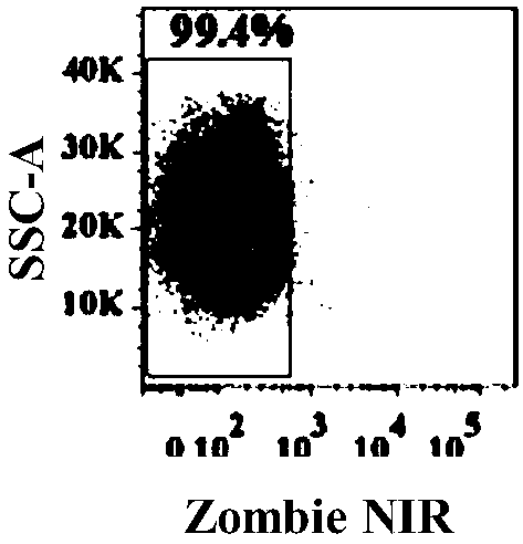
[0105] 2) Add 1mL cell staining buffer to each tube, centrifuge at 200g for 6min, and discard the supernatant; the obtained human peripheral blood mononuclear cell population after removing dead cells is as follows: figure 2 shown;

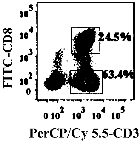
[0106] 3) Add 100 μL of cell staining buffer to each tube to resuspend the cells, then add PerCP / Cy 5.5-labeled anti-human CD3, FITC-labeled anti-human CD8, PE-labeled anti-human CD25, BrilliantViolet 421-labeled anti-human TGF-β and BrilliantViolet 510-labeled anti-human CD127 antibody 5uL each, or PerCP / Cy 5.5-labeled anti-human CD3, FITC-labeled anti-human CD8, BrilliantViolet 510-labeled anti-human CD25, PE / Cy7-labeled anti-human CD278 (ICOS) and PE Incubate 5uL of each labeled anti-human CCR6 antibody at room temperature in the d...


PUM

PropertyMeasurementUnit
osmolarityaaaaaaaaaa

Abstract

The invention provides a kit for detecting human regulatory T cell subtypes and a detection method and belongs to the technical field of cell subtype detection. The provided kit comprises components as follows: a blood diluent, a mononuclear cell separation medium, a cell culture fluid, a lymphocyte activation solution, dead cell removal dyes, an FcR blocking agent, a fluorescence labeled antibodyresisting human cell surface labeling molecules, a fluorescence labeled antibody resisting human intracellular molecules, a PBS buffer solution, lipopolysaccharide, a washing buffer solution, a celldyeing buffer solution, a cell fixing solution and a permeable membrane lotion. During detection, firstly, mononuclear cells in whole blood are separated, then, mononuclear cells of human peripheral blood are stimulated, finally, a fluorescent antibody is stained, and the regulatory T cell subtypes can be detected. By use of the kit and the detection method, 2-6 regulatory T cell subtypes can be simply and rapidly distinguished.

Description

technical field [0001] The invention belongs to the technical field of cell subtype detection, and in particular relates to a kit and a detection method for detecting human regulatory T cell subtype. Background technique [0002] In the mid-1990s, it was proposed that CD4 + Suppressive T cells, namely regulatory T (Treg) cells, can suppress the immune damage effect of effector T cells such as Th cells on the body. At present, more and more data show that there are several different subtypes of Treg in the human body, and their roles in the immune system are different, which makes the role of Treg more complicated. [0003] Naive T cells can differentiate into a variety of regulatory T (Treg) cells under different conditions, and at least seven Treg cell subtypes have been identified: (1) nTregs: CD4 that secretes IL-10 and TGF-β + CD25 + Foxp3 + T cells are the most numerous Treg cell subtype; (2) iTregs: Foreign antigens can stimulate peripheral blood naive CD4 + T cel...

Claims


Application Information

Patent Timeline
Patent Type & Authority Applications(China)
IPC IPC(8): G01N33/533G01N33/50
CPCG01N33/505G01N33/533G01N2333/4704G01N33/56972G01N1/30G01N2333/4703G01N2333/495G01N2333/54G01N2333/5428G01N2333/7051G01N2333/70517G01N2333/715
Inventor 何韶衡何萍
Owner 沈阳汇敏源生物科技有限责任公司
Why Eureka
Try Eureka