
Three-dimensional live cell super-resolution microscopy imaging method and device based on evanescent wave illumination

A microscopic imaging and super-resolution image technology, applied in the field of optical super-resolution microscopic imaging, can solve the problems that cannot be used to observe the three-dimensional fine structure of the sample, lose the total internal reflection fluorescence microscope, and it is difficult to break through the half-wavelength order. Achieve three-dimensional fast super-resolution imaging, strong applicability, and simple structure

Active Publication Date: 2020-08-18
ZHEJIANG UNIV
View PDF7 Cites 0 Cited by
  • Summary
  • Abstract
  • Description
  • Claims
  • Application Information

AI Technical Summary

Problems solved by technology

However, this method needs to obtain dozens of original images to obtain tens of nanometers of axial resolution, which to some extent loses the greatest advantage of total internal reflection fluorescence microscopy, and limits its application in real-time imaging of living cells.
[0006] On the other hand, the total internal reflection fluorescence microscope only improves the axial resolution of the system, and its lateral resolution is still limited by the diffraction limit. It is difficult to break through the half-wavelength order and cannot be used to observe the three-dimensional fine structure of the sample.

Method used


Image

  • Three-dimensional live cell super-resolution microscopy imaging method and device based on evanescent wave illumination
  • Three-dimensional live cell super-resolution microscopy imaging method and device based on evanescent wave illumination
  • Three-dimensional live cell super-resolution microscopy imaging method and device based on evanescent wave illumination

Examples


Embodiment

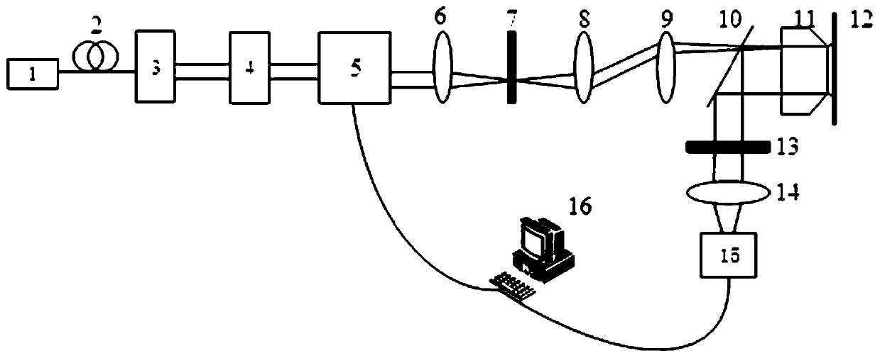
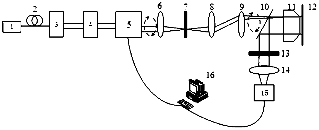
[0045] see figure 2 with image 3 , the three-dimensional super-resolution microscopic imaging device of this embodiment includes an excitation optical path module, a detection optical path module, and a processing module.

[0046]The excitation optical path module includes a laser 1 , a single-mode polarization-maintaining fiber 2 , a quarter-wave plate 3 , a half-wave plate 4 , a scanning galvanometer 5 , a lens group, a polarization rotator 7 and a microscope objective 11 . The laser 1 is used to generate an illuminating laser beam, and the generated laser beam passes through a single-mode polarization-maintaining fiber 2 to generate a single-mode laser beam, and the polarization direction of the generated single-mode laser beam is controlled by a quarter-wave plate 3 and a half-wave plate 4 . By rotating the quarter-wave plate 3, the laser beam can be converted between circularly polarized light and linearly polarized light, and by rotating the half-wave plate 4, the pol...


PUM


Abstract

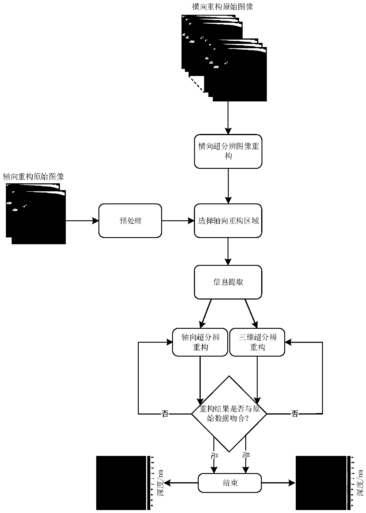
The invention discloses a three-dimensional living cell super-resolution microscopic imaging method and device based on evanescent wave illumination, and belongs to the field of optical super-resolution microscopic imaging. The method comprises the following implementation steps: acquiring a series of transverse super-resolution reconstructed original images by illuminating a sample with incidentlight at a single angle larger than the critical angle of total reflection, and reconstructing all transverse super-resolution original images to obtain a transverse super-resolution image of the sample; illuminating the sample with incident light larger than the critical angle of total reflection at any two incident angles, and performing annular scanning at the same time to obtain two corresponding axial super-resolution reconstructed original images; performing background subtraction preprocessing on the axial super-resolution reconstructed original images, performing information extractionby taking a single axial reconstructed original image and the transverse super-resolution image as templates respectively at the same time, and performing axial super-resolution reconstruction and three-dimensional super-resolution reconstruction on the extracted images respectively to obtain an axial super-resolution image and a three-dimensional super-resolution image.

Description

technical field [0001] The invention relates to the field of optical super-resolution microscopic imaging, in particular to a three-dimensional live cell super-resolution microscopic imaging method and device based on evanescent wave illumination. Background technique [0002] Observing and studying cell structure and function is a branch of biology. Cells are the basic units that make up the shape and function of organisms, and they themselves are composed of many parts. The research on the structure not only needs to know which parts it is composed of, but also needs to further clarify the composition of each part. Correspondingly, with regard to function, it is not only necessary to know the function of the cell as a whole, but also to understand the functional interrelationships of various parts. For a long time, limited by the optical diffraction limit, traditional optical microscopes cannot distinguish the detailed information of objects in the lateral range of 200nm...

Claims


Application Information

Patent Timeline
Patent Type & Authority Patents(China)
IPC IPC(8): G01N21/64
CPCG01N21/6486
Inventor 匡翠方刘文杰刘旭张克奇毛磊
Owner ZHEJIANG UNIV
Learn More