Method and kit for detecting blood-drug concentration of PD-1 antibody

A technology of PD-1 and blood drug concentration, applied in the direction of measuring devices, instruments, scientific instruments, etc., can solve the problem of high detection background and achieve good sensitivity

Active Publication Date: 2019-03-05
ACROBIOSYSTEMS INC
View PDF8 Cites 3 Cited by
  • Summary
  • Abstract
  • Description
  • Claims
  • Application Information

AI Technical Summary

Problems solved by technology

[0006] Most of the preclinical and clinical pharmacokinetic studies of the traditional ELISA for the detection of PD-1 antibodies are indirect methods. As we all kno

Method used


Image

  • Method and kit for detecting blood-drug concentration of PD-1 antibody
  • Method and kit for detecting blood-drug concentration of PD-1 antibody
  • Method and kit for detecting blood-drug concentration of PD-1 antibody

Examples


Embodiment 1

[0041] 1. Material

[0042] 1.1 Drugs and reagents: PD-1, biotinylated anti-PD-1 (biotin-labeled PD-1 antibody); standard: PD-1 monoclonal antibody, 200mg; coating solution: 1.6g Na 2 CO 3 , 2.9g NaHCO 3 , 0.5g NaN 3 , Add double-distilled water to 1L, adjust the pH to 9.6, filter, aliquot and store at 4℃; PBS: 0.2722g KH2PO4, 3.58g Na2HPO4.12H2O, 8.063g NaCl, 0.2066g ​​KCl, add double-distilled water to 1L, Adjust the pH to 7.4, filter, aliquot, and store at 4°C; washing solution (PBST): add 0.05% Tween 20 to PBS. Shake well before use; blocking solution: PBST solution containing 2% BSA; sample diluent: PBST solution containing 0.5% BSA; antibody diluent: PBST solution containing 0.5% BSA; antibody: Streptavidin Protein, HRPconjugate (21126) form Thermo, use ratio 1:10000; blank serum: purchased from Shunran (Shanghai) Biotechnology Co., Ltd. Wherein, the biotinylated anti-PD-1 is prepared by the following method: the antibody concentration is controlled to between 0.8mg / ml, an...

Embodiment 2

[0095] 1. Establish the quality inspection standard of each component stock solution

[0096] According to 1.4 ELISA operation steps, the quality inspection of each component is performed separately. The quality inspection standard of each component is that the experimental data deviation (RSD) of different days is less than 20%. The stock solution that has passed the quality inspection can be used as the component in the kit. The experimental results show that the deviation of the IC50 value in different days does not exceed 20% (Table 10).

[0097] Table 10 The deviation of the stock solution components in different days

[0098]

[0099] 2. Lyophilization of kit components

[0100] The freeze-drying process is: pre-freezing temperature is -60℃, lasts 6 hours, cold trap temperature is -70℃, vacuum degree is guaranteed below 10Pa, drying time is 30 hours, and the freeze-dried product has a full appearance.

[0101] 3. Quality inspection of the kit

[0102] After lyophilization, each co...

Embodiment 3

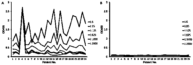
[0122] The method in this example is a general method that can detect multiple PD-1 antibodies (provided by different manufacturers) under research and on the market. The specific experiment is as follows, according to 1.4 ELISA procedure to carry out various antibody verification, the result shows ( figure 2 ), different antibody drugs may use this method for serum drug content determination.


PUM


Abstract

The invention provides a method and a kit for detecting the blood-drug concentration of a PD-1 antibody. With the method provided by the invention, the concentration of the PD-1 antibody is detected by competition of a biotin-labeled PD-1 neutralizing antibody with the PD-1 antibody in a to-be-detected sample through a competitive ELISA method, wherein the biotin-labeled PD-1 neutralizing antibodycompetes with the PD-1 antibody in serum in binding to an antigen immobilized on an elisa plate, and horseradish peroxidase labeled streptavidin is used as a detection antibody to detect the biotin-labeled PD-1 neutralizing antibody, so the PD-1 antibody in the serum is quantified. The method provided by the invention has the advantages of good sensitivity, precision, repeated freeze-thawing stability, room-temperature storage stability, specificity, etc., and facilitates forming a commercial kit. The kit provided by the invention has the advantages of low background, good precision, batch-to-batch consistency, stability, etc.

Description

Technical field [0001] The present invention relates to a method and kit for detecting the plasma concentration of PD-1 antibody, specifically, to an enzyme-linked immunosorbent assay (ELISA) for measuring the concentration of PD-1 antibody in serum and the reagents used box. Background technique [0002] In recent years, monoclonal antibody drugs designed based on immune checkpoint proteins such as programmed cell apoptosis protein-1 (PD-1) and its ligands (PD-L1 / L2) have been widely used as an important immune regulatory strategy for tumor suppression. Currently, FDA-approved monoclonal antibody drugs against PD-1 target include Merck's Keytruda (pembrolizumab) and Bristol-Myers Squibb Opdivo (nivolumab), but they have not yet been marketed in my country. In addition, anti-PD-1 related monoclonal antibody drugs are also declared by many domestic pharmaceutical manufacturers, but the current clinical research of PD-1 monoclonal antibody drugs is initiated by multiple sponsors, ...

Claims


Application Information

Patent Timeline
IPC IPC(8): G01N33/558
CPCG01N33/558
Inventor 陈宜顶葛平菊平志光
Owner ACROBIOSYSTEMS INC
Why Eureka
Try Eureka