
Controllable large-scale preparation method of NK cell exosomes

A technology of NK cells and phosphate buffer solution, which is applied in the field of controllable and large-scale preparation of NK cell exosomes, can solve the problems of poor removal of impurity proteins and low yield, and achieve high scientific research value and uniform particle size distribution , the effect of high safety factor

Inactive Publication Date: 2019-03-08
SHANGHAI JIAO TONG UNIV
View PDF3 Cites 0 Cited by
  • Summary
  • Abstract
  • Description
  • Claims
  • Application Information

AI Technical Summary

Problems solved by technology

The purity of exosomes obtained by traditional ultra-high-speed centrifugation is acceptable, but its yield is not high, and ultrafiltration, dialysis and other methods designed according to the principle of exosome size cannot remove impurity proteins that are consistent with the size of exosomes, and polyethylene glycol Both the (polyethylene glycol, PEG) precipitation method and the immunomagnetic bead method introduce exogenous substances that are not suitable for clinical treatment to varying degrees.

Method used


Image

  • Controllable large-scale preparation method of NK cell exosomes
  • Controllable large-scale preparation method of NK cell exosomes
  • Controllable large-scale preparation method of NK cell exosomes

Examples


Embodiment 1

[0027] (1) Dilute the collected NK cell culture supernatant with phosphate buffered saline at 4°C, centrifuge at 3,000 rpm for 8 min, precipitate into cells, remove the precipitate, and keep the supernatant;

[0028] (2) Centrifuge the supernatant obtained in step (1) at 9,000 rpm for 15 minutes at 4°C, retain the supernatant, and discard the precipitate;

[0029] (3) Dilute the supernatant obtained in step (2) with phosphate buffer at 4°C, continue to centrifuge at 3,500 rpm for 5 min with a 110kDa ultrafiltration tube to remove small molecular impurities and concentrate the cell supernatant , to reduce the workload and cost of the subsequent ultra-high-speed centrifugation;

[0030] (4) Centrifuge the concentrated supernatant obtained in step (3) at a speed of 100,000 g at 4°C for 60 min. After the centrifugation, discard the supernatant and retain the precipitate;

[0031] (5) Resuspend the precipitate obtained in step (4) with phosphate buffer and transfer it to a dialysi...

Embodiment 2

[0037] (1) Dilute the collected NK cell culture supernatant with phosphate buffered saline at 4°C, centrifuge at 3,000rpm for 9min, precipitate into cells, remove the precipitate, and keep the supernatant,

[0038] (2) Centrifuge the supernatant obtained in step (1) at a speed of 10,000 rpm for 20 minutes at 4°C, retain the supernatant, and discard the precipitate;

[0039] (3) Dilute the supernatant obtained in step (2) with phosphate buffer at 4°C, continue to centrifuge at 3,500 rpm for 5 min with a 110kDa ultrafiltration tube to remove small molecular impurities and concentrate the cell supernatant , to reduce the workload and cost of the subsequent ultra-high-speed centrifugation.

[0040] (4) The concentrated supernatant obtained in step (3) was centrifuged at 100,000 g for 60 min at 4°C. After centrifugation, the supernatant was discarded and the precipitate was retained.

[0041] (5) The precipitate obtained in step (4) was resuspended in phosphate buffer and transfer...

Embodiment 3

[0043] (1) Dilute the collected NK cell culture supernatant with phosphate buffered saline at 4°C, centrifuge at 3,000rpm for 8min, precipitate into cells, remove the precipitate, and keep the supernatant,

[0044] (2) Centrifuge the supernatant obtained in step (1) at 10,000 rpm for 10 min at 4°C, retain the supernatant, and discard the precipitate;

[0045] (3) Dilute the supernatant obtained in step (2) with phosphate buffer at 4°C, continue centrifuging at 3,000 rpm for 10 min with a 110kDa ultrafiltration tube to remove small molecule impurities and concentrate the cell supernatant , to reduce the workload and cost of the subsequent ultra-high-speed centrifugation.

[0046] (4) The concentrated supernatant obtained in step (3) was centrifuged at 100,000 g for 60 min at 4°C. After centrifugation, the supernatant was discarded and the precipitate was retained.

[0047] (5) The precipitate obtained in step (4) was resuspended in phosphate buffer and transferred to a dialysi...


PUM


Abstract

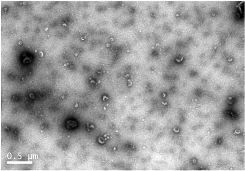
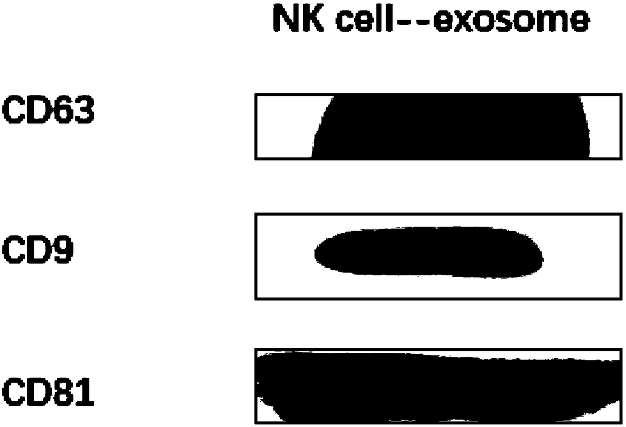
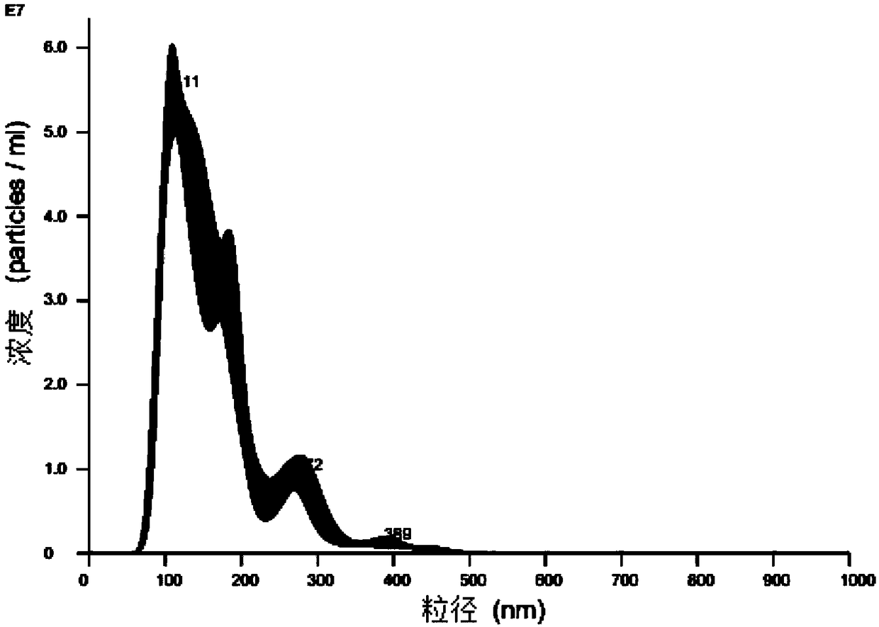
The invention relates to a controllable large-scale preparation method of NK cell exosomes. The method includes the steps: (1) collecting NK cell culture medium liquid supernatant, diluting the liquidsupernatant by phosphate buffer solution to obtain dilution solution; (2) sequentially centrifuging the dilution solution at low rotating speed and high rotating speed, and taking liquid supernatantand removing precipitate after centrifuging once; (3) placing centrifuged liquid supernatant into an ultra-filtration tube to centrifuge the liquid supernatant, taking liquid supernatant, centrifugingacquired concentrated liquid supernatant at ultra-high speed, and taking precipitate after ultra-high speed centrifugation; (4) placing the precipitate acquired after ultra-high speed centrifugationin the step (3) into phosphate buffer solution, and transferring mixture into a dialysis bag to perform dialysis to obtain NK cell exosomes. Compared with the prior art, particle diameters are uniformly distributed, the surfaces of the NK cell exosomes have iconic protein CD9, CD81 and CD63, the method overcomes the shortcomings of low purity, yield and characterization protein expression of an existing preparation technique and the like, and the method can be directly used for subsequent detection and is high in safety coefficient.

Description

technical field [0001] The invention relates to the field of biotechnology, in particular to a controllable and large-scale preparation method of NK cell exosomes. Background technique [0002] Exosomes are 30-150nm "cup holder-shaped" phospholipid bilayer vesicles secreted by many types of cells, with a density of 1.13-1.19g / mL, carrying a variety of proteins, lipids and nucleic acids, with complex functions, involved in immune regulation, Regulate extracellular matrix remodeling, activate cell signaling pathways, etc. In cancer research, most previous studies have explored the exosomes released by cancer cells, but little is known about the function of exosomes released by immune cells. NK cells have a natural and rapid immune effect on metastatic and hematological malignancies, and the anti-tumor properties of NK cells have been in the clinical trial stage. However, live cell therapy carries inherent risks: microvascular occlusion leading to pulmonary embolism and death...

Claims


Application Information

Patent Timeline
IPC IPC(8): C12N5/0783
CPCC12N5/0646
Inventor 崔大祥潘少君张倩章阿敏黄志成
Owner SHANGHAI JIAO TONG UNIV
Learn More