Preparation method of antibody orientation modified fluorescent microsphere probe and application thereof in immunochromatography
A fluorescent microsphere and antibody technology, applied in the field of nanomaterials and immunoassays, can solve the problems of antibody shedding and probe activity reduction, and achieve the effects of avoiding occupation, improving sensitivity and improving efficiency.
- Summary
- Abstract
- Description
- Claims
- Application Information
AI Technical Summary
Problems solved by technology
Method used
Image
Examples
Embodiment 1
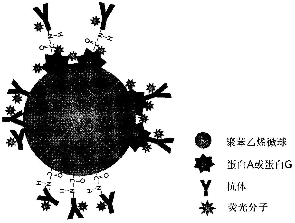
[0029] Example 1-Preparation of fluorescent microsphere probes targeting cTnI (cardiac troponin I) antibody orientation modification
[0030] Protein G was coupled to EDC / NHS activated carboxyl microspheres, and then incubated with anti-cTnI antibody and fixed with EDC, and finally coupled with fluorescent molecules.
[0031] Proceed as follows:
[0032] 1) Disperse 1mg of carboxyl microspheres with a particle size of 150nm in 1mL pH6 PBS buffer, add 50μg EDC and 50μg NHS, and activate at 25°C for 30 minutes;
[0033] 2) Centrifuge to remove excess cross-linking agent, disperse the precipitate in 1mL pH7 PBS buffer, add 200μg protein G, and incubate at 25°C for 1h to couple protein G to the microspheres;
[0034] 3) Wash by centrifugation, disperse the precipitate in 200μL pH7 PBS buffer, add 200μg of anti-cTnI monoclonal antibody (mouse source), and incubate at 25°C for 1h;
[0035] 4) Add 50μg EDC to the reaction system, and continue coupling at 25°C for 2h, so that the antibody is fix...
Embodiment 2
[0039] Example 2-Preparation of lateral immunochromatographic fluorescent test strips for detecting cTnI
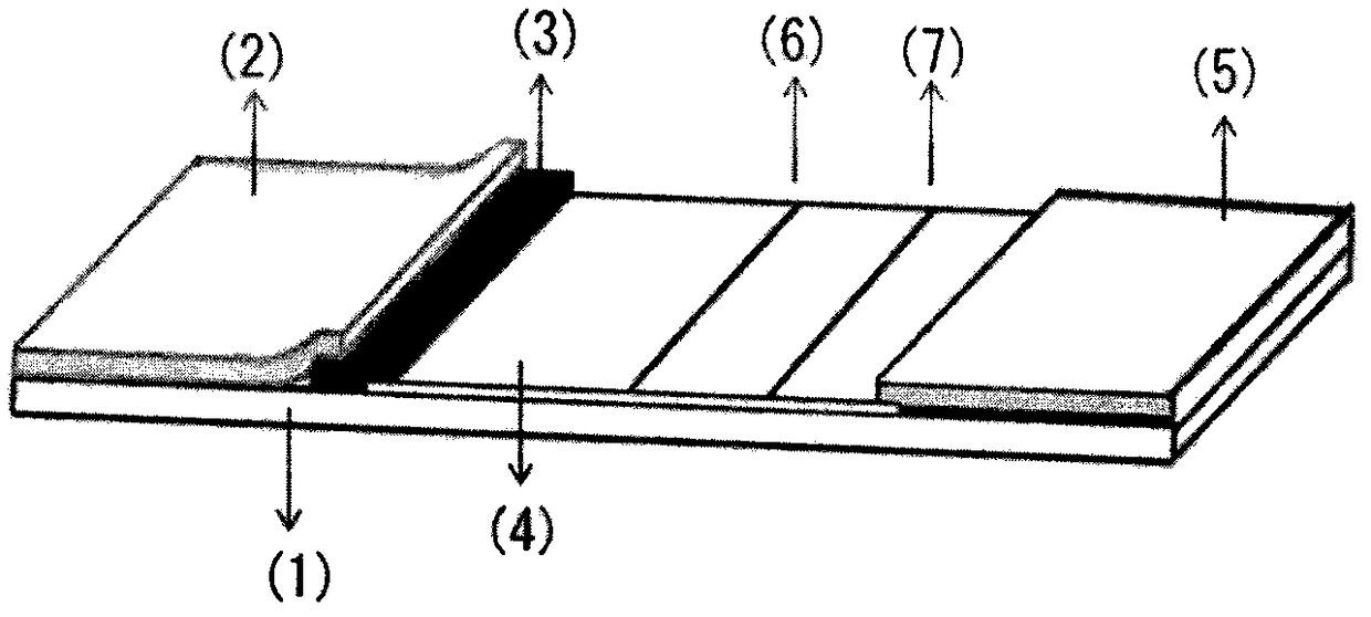
[0040] The basic structure of the test strip is as follows figure 1 Shown. It is composed of a PVC bottom plate (1), a sample pad (2), a bonding pad (3), a nitrocellulose membrane (4) and absorbent paper (5) which are sequentially overlapped on the bottom plate (1). The adjacent components of each overlapping part need to overlap by about 2mm to ensure the smooth chromatography of the sample on the test strip. The assembled and chopped test strips need to be put into a card case, sealed in a tin foil bag containing a desiccant, and stored in a cool, dry environment.
[0041] Specific steps are as follows:
[0042] 1) Treat sample pads and binding pads: add 0.5% inert protein (such as BSA), 0.05% (such as Tween-20) and 0.05% high molecular polymer (such as PEG2K) to Tris buffer, and mix well. Adjust the pH to 7-8 to obtain the treatment solution; each mat (specification 20cm×3...
Embodiment 3
[0052] Example 3-Detection of cTnI in samples by lateral immunochromatographic test strips
[0053] Specific steps are as follows:
[0054] 1) Restore the serum, plasma or whole blood sample to room temperature;
[0055] 2) Take the test strip out of the tin foil bag;
[0056] 3) Take 100μL of sample and drop it into the sample hole;
[0057] After 15 minutes of chromatography, insert the test strip into the fluorescence quantitative immunoassay analyzer to read the test results.
PUM
 Abstract
Description
Claims
Application Information
 - R&D Engineer
- R&D Manager
- IP Professional
- Industry Leading Data Capabilities
- Powerful AI technology
- Patent DNA Extraction
Browse by: Latest US Patents, China's latest patents, Technical Efficacy Thesaurus, Application Domain, Technology Topic, Popular Technical Reports.
© 2024 PatSnap. All rights reserved.Legal|Privacy policy|Modern Slavery Act Transparency Statement|Sitemap|About US| Contact US: help@patsnap.com


