Rapid tissue transparent processing method based on water-soluble reagent and application thereof
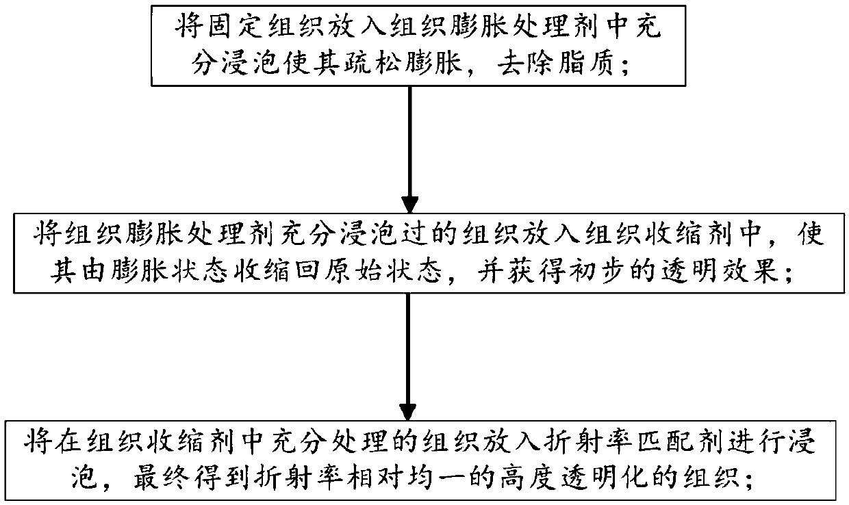
A processing method and water-soluble technology, applied in the field of biomedical optical imaging, can solve the problems of limiting the application of light-transparency methods, slow transparent speed, limited transparent effect, etc., and achieve the effect of fast transparent speed, improved imaging depth, and short transparent time.
- Summary
- Abstract
- Description
- Claims
- Application Information
AI Technical Summary
Problems solved by technology
Method used
Image
Examples
Embodiment 1
[0042] The biological tissue of the embodiment of the present invention is taken from the brain tissue of a C57 mouse, and its 2mm slices are processed through the transparent treatment method of the embodiment 1 of the present invention, which specifically includes the following steps:
[0043]First, soak the thin slices in the tissue swelling agent (10% m-xylylenediamine, 5% sorbitol) for 4-6 hours; then soak the thin slices in the tissue shrinking agent (m-xylylenediamine 30%) %, sorbitol 20%, phosphate buffer saline 0.03M), processing time 2-3h; finally the section is soaked in the refractive index matching agent (m-xylylenediamine 40%, sorbitol 40%), processing time 1- 2h;
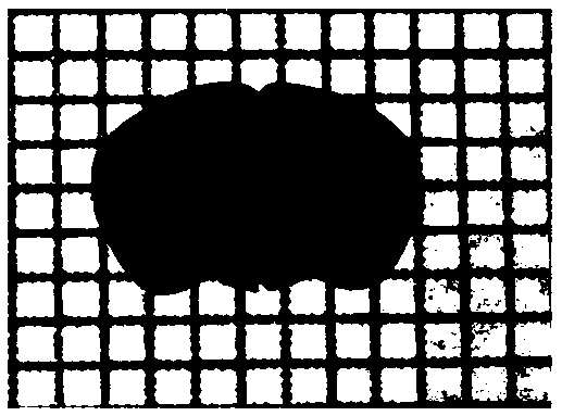
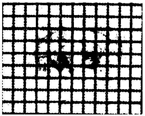
[0044] Figure 2a-2b Visual images taken before and after light-transparent treatment of isolated mouse 2mm brain slices are given. in Figure 2a For the situation before the transparent treatment, due to the turbidity of the tissue, the lines below the tissue are completely covered; Figure 2b Fo...
Embodiment 2
[0046] The biological tissue of the embodiment of the present invention is taken from the brain tissue of Thy-1EGFP and Cx3cr1-EGFP mice, and its 2mm slices are processed through the transparent processing method of the embodiment 1 of the present invention, which specifically includes the following steps:
[0047] First, soak the thin slices in the tissue swelling agent (10% m-xylylenediamine, 5% sorbitol) for 4-6 hours; then soak the thin slices in the tissue shrinking agent (m-xylylenediamine 30%) %, sorbitol 20%, phosphate buffer saline 0.05M), processing time 2-3h; finally the section is soaked in the refractive index matching agent (m-xylylenediamine 40%, sorbitol 40%), processing time 1- 2h; immerse the transparent 2mm brain slice in the refractive index matching agent for confocal fluorescence imaging.
[0048] Figures 3a-3c Fluorescence images of cortical pyramidal neurons and microglia taken before and after the light-transparent treatment of the mouse brain slices...
Embodiment 3
[0050] The biological tissue of the embodiment of the present invention is taken from various organs of C57 mice, and the whole organ is processed through the transparent treatment method of the embodiment 3 of the present invention, which specifically includes the following steps:
[0051] First, soak various organs in the tissue swelling treatment agent (m-xylylenediamine 20%, sorbitol 10%). The soaking time is respectively according to the tissue type: mouse whole brain 24-36h, mouse heart 12-24h, Liver 24-36h, kidney 12-24h, lung 12-24h, spleen 12-24h, stomach 6-12h; then soak each organ in tissue contraction agent (40% m-xylylenediamine, 30% sorbitol, phosphoric acid Salt buffer 0.01M), the soaking time according to the tissue type is: mouse whole brain 8-12h, mouse heart 4-8h, liver 8-12h, kidney 8-12h, lung 8-12h, spleen 4-8h, Stomach 4-8h; Finally, each organ is soaked in the refractive index matching agent (m-xylylenediamine 50%, sorbitol 40%), mouse whole brain 8-12h...
PUM
 Abstract
Description
Claims
Application Information
 - R&D
- Intellectual Property
- Life Sciences
- Materials
- Tech Scout
- Unparalleled Data Quality
- Higher Quality Content
- 60% Fewer Hallucinations
Browse by: Latest US Patents, China's latest patents, Technical Efficacy Thesaurus, Application Domain, Technology Topic, Popular Technical Reports.
© 2025 PatSnap. All rights reserved.Legal|Privacy policy|Modern Slavery Act Transparency Statement|Sitemap|About US| Contact US: help@patsnap.com


