Fluorescent probe for detecting N-acetyl-beta-D-glucosaminidase and application thereof

A technology of glucosamine and glucosamine, which is applied in the field of fluorescent probes for detecting N-acetyl-β-D-glucosaminidase, can solve the problems of complicated operation and low sensitivity, achieve high sensitivity, low detection cost, good distraction effect

Active Publication Date: 2019-05-31
DALIAN MEDICAL UNIVERSITY
View PDF3 Cites 4 Cited by
  • Summary
  • Abstract
  • Description
  • Claims
  • Application Information

AI Technical Summary

Problems solved by technology

At present, the detection methods used in clinical practice have problems such as low sensitivity and complicated operation. Therefore, it is necessary to develop a specific N-acetyl-β-D-glucosaminidase with high sensitivity and strong anti-environmental interference ability for in vivo and in vitro detection. Substrates for fluorescent fluorescent probes, and the establishment of high-sensitivity scientific detection methods, have important application value in clinical diagnosis, drug development and rational use

Method used


Image

  • Fluorescent probe for detecting N-acetyl-beta-D-glucosaminidase and application thereof
  • Fluorescent probe for detecting N-acetyl-beta-D-glucosaminidase and application thereof
  • Fluorescent probe for detecting N-acetyl-beta-D-glucosaminidase and application thereof

Examples


Embodiment 1

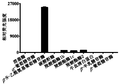
[0025] Example 1. In vitro determination of the selectivity of different hydrolases

[0026] (1) Prepare 99 µL in vitro metabolic reaction system in advance, including pH 7.4 phosphate buffer (100 mM), different kinds of hydrolytic enzymes (0.1 mg / mL), at 37 o Pre-incubation with shaking for 3 minutes under C condition;

[0027] (2) Add 1 µL of DDAG at a concentration of 1 mM (final concentration 10 μM) to the reaction system to initiate the reaction;

[0028] (3) After 30 minutes, add 50 µL of glacial acetonitrile and shake vigorously to terminate the reaction;

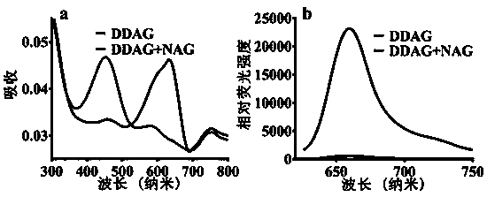
[0029] (4) Use a high-speed refrigerated centrifuge at 4 o C. 20,000 × g Under the condition of high-speed centrifugation for 20 minutes, the supernatant was taken for fluorescence detection (DDAG: Ex=608 nm, Em=660 nm). The results showed that only N-acetyl-β-D-glucosaminidase (NAG) catalyzed the reaction, and the reaction rate was much higher than that of other hydrolytic enzymes, indicating that N-acetyl-β-D-g...

Embodiment 2

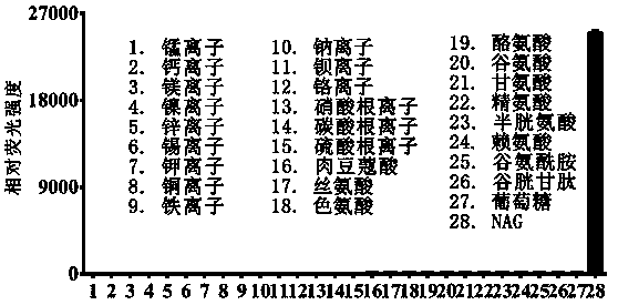
[0030] Example 2. In vitro determination of the selectivity of different hydrolases

[0031] (1) Prepare 99 µL in vitro metabolic reaction system in advance, including pH 7.4 phosphate buffer (100 mM), different kinds of hydrolytic enzymes (0.1 mg / mL), at 37 o Pre-incubation with shaking for 3 minutes under C condition;

[0032] (2) Add 1 µL of DDAG at a concentration of 1 mM (final concentration 10 μM) to the reaction system to initiate the reaction;

[0033] (3) After 30 minutes, add 50 µL of glacial acetonitrile and shake vigorously to terminate the reaction;

[0034] (4) Use a high-speed refrigerated centrifuge at 4 o C. 20,000 × g Under the condition of high-speed centrifugation for 20 minutes, the supernatant was taken for fluorescence detection (DDAG: Ex=608 nm, Em=660 nm). The results showed that only N-acetyl-β-D-glucosaminidase (NAG) catalyzed the reaction, and other ions and amino acids had no effect on the fluorescence intensity of the probe, indicating that N-...

Embodiment 3

[0035] Example 3. Linearity study of N-acetyl-β-D-glucosaminidase catalyzed DDAG probe reaction

[0036] (1) Prepare 99 µL in vitro metabolic reaction system in advance, including pH 7.4 phosphate buffer (100 mM), N-acetyl-β-D-glucosaminidase (0-3 μg / mL), at 37 o Pre-incubation with shaking for 3 minutes under C condition;

[0037] (2) Add 1 µL of NHPO with a concentration of 1 mM (final concentration 10 μM) to the reaction system to initiate the reaction;

[0038] (3) After 30 minutes, add 50 µL of glacial acetonitrile and shake vigorously to terminate the reaction;

[0039] (4) Use a high-speed refrigerated centrifuge at 4 o C. 20,000 × g Under the condition of high-speed centrifugation for 20 minutes, the supernatant was taken for fluorescence detection (DDAG: Ex=608 nm, Em=660 nm). The results showed that the probe reaction of N-acetyl-β-D-glucosaminidase (NAG) catalyzed DDAG showed a good enzyme linear relationship in the range of 0-3 μg / mL, r 2Value is 0.9945, illus...


PUM


Abstract

The invention discloses a fluorescent probe for detecting N-acetyl-beta-D-glucosaminidase and application thereof, and belongs to the technical field of biological medicines. The fluorescent probe canbe used for determining the enzymatic activity of N-acetyl-beta-D-glucosaminidase in different biological systems. The activity of N-acetyl-beta-D-glucosaminidasein each biological sample is determined by quantitatively detecting the production amount of the aglycone metabolites in unit time, with DDAG as a specific probe reaction substrate, in vitro reaction system of N-acetyl-beta-D-glucosaminidase as means andthe hydrolysis reaction of beta-glucuronideas a probe reaction. The fluorescent probecan be used for the qualitative and quantitative determination of the N-acetyl-beta-D-glucosaminidaseactivity in human and animal tissue samples of different individual sources, different species cells and cell preparations and various plants and microorganisms, and can achieve theevaluation of the ability of N-acetyl-beta-D-glucosaminidase to treat drugs, and the development and screening of N-acetyl-beta-D-glucosaminidase inhibitors.

Description

technical field [0001] The invention belongs to the technical field of biomedicine, and in particular relates to a fluorescent probe for detecting N-acetyl-β-D-glucosaminidase and its application. Background technique [0002] N-acetyl-β-D-glucosaminidase (N-acetyl-β-D-glucosaminidase, NAG) is an acidic hydrolase of polymer glycoproteins, widely present in lysosomes of various tissues in the body, especially It is most abundant in the kidney. When renal tubules are damaged or slightly damaged in the early stage, the filtration and absorption of NAG by renal tubular cells are changed, which leads to a significant increase in the activity of NAG in urine. [0003] NAG in urine is a sensitive and specific indicator reflecting renal tubular damage, and its activity determination is widely used in the clinical monitoring of various renal diseases. At present, the detection methods used in clinical practice have problems such as low sensitivity and complicated operation. Therefo...

Claims


Application Information

Patent Timeline
IPC IPC(8): C07H17/02C09K11/06G01N33/58
Inventor 马骁驰冯磊田象阁宁静王超于振龙霍晓奎孙成鹏
Owner DALIAN MEDICAL UNIVERSITY
Why Eureka
Try Eureka