Improved fluorescent protein transgenic mouse tissue fluorescence imaging method

A transgenic mouse, fluorescent protein technology, applied in fluorescence/phosphorescence, material excitation analysis, etc., can solve the problems of difficult localization of GFP fluorescent protein, loss of tissue, hindering popularization and application, etc., to make up for the loss of signal in tissue slices, to avoid deterioration and fragmentation , the effect of improving the efficiency of nuclear staining

Active Publication Date: 2019-06-07
WUHAN UNIV
View PDF2 Cites 2 Cited by
  • Summary
  • Abstract
  • Description
  • Claims
  • Application Information

AI Technical Summary

Problems solved by technology

The signal obtained by this method depends largely on the efficiency of the immunofluorescence reaction, which is affected by many factors, such as antibody titer and reaction system stability; the photos obtained by this method are discontinuous 2D planes The image cannot be used to construct a 3D stereoscopic image; and some tissues will inevitably be lost during the slicing process, resulting in signal loss
[0005] (3) Transgenic mouse hybridization and confocal imaging technology: In addition to the above two commonly used methods, in order to obtain 3D images of the cellular and subcellular localization of genes in tissues, some researchers also tried to culture GFP transgenic mouse tooth germ tissue Directly placed under the confocal microscope to take imag

Method used


Image

  • Improved fluorescent protein transgenic mouse tissue fluorescence imaging method
  • Improved fluorescent protein transgenic mouse tissue fluorescence imaging method

Examples


Embodiment 1

[0038] The features and advantages of the present invention can be further understood through the following detailed description in conjunction with the accompanying drawings. The examples provided are only illustrative of the method of the present invention and do not limit the rest of the present disclosure in any way. [Example 1] The process of determining the fixed temperature, fixed time, Triton concentration, nuclear staining dye and nuclear staining time in the fluorescence imaging method of GFP transgenic mouse tooth germ tissue is as follows:

[0039] (1) Fixed temperature and fixed time: Fix the incisor terminal tissue pieces of P2GFP transgenic mice in 4% paraformaldehyde at room temperature or 4 degrees for 1 minute, 2 minutes, 5 minutes, 10 minutes, 30 minutes, 60 minutes respectively . In the subsequent counterstaining and imaging process, the tissue integrity and GFP fluorescence signal intensity are the best when the tissue is fixed at 4 degrees for 5 minutes;...

Embodiment 2

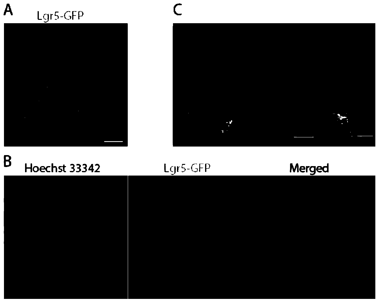
[0042] [Example 2] Confocal fluorescence imaging of incisor tooth germ tissues of Lgr5-GFP transgenic mice

[0043] 1. Materials and methods

[0044] 1.1 Materials

[0045] Animals: one litter (6-8) of Lgr5-GFP transgenic mice 2 days after birth (P2);

[0046] Reagents: 1×PBS, 1×PBS-0.5% Triton, 10mg / mL Hoechst 33342, Vestsheild;

[0047] Consumables and instruments: glass slides, coverslips, confocal microscope.

[0048] 1.2 Method

[0049] (1) Dissect and obtain the incisor terminal tissue pieces of Lgr5-GFP transgenic P2 mice, rinse slowly with shaking in 1×PBS at room temperature for 5 minutes×2 times, soak in 4% paraformaldehyde, and place at 4°C for slow shaking , fix for 5 minutes, after fixation, slowly shake and rinse in 1×PBS at room temperature for 5 minutes×2 times;

[0050] (2) Soak the fixed tissue block in 1×PBS-0.5% Triton, shake slowly and rinse for 5 minutes×2 times;

[0051] (3) Soak the above-treated tooth germ tissue block in the fluorescent dye Hoes...

Embodiment 3

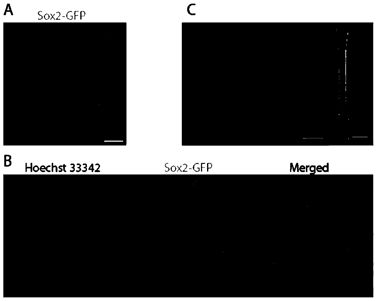
[0057] [Example 3] Confocal fluorescence imaging of incisor tooth germ tissue in Sox2-GFP transgenic mice

[0058] 1. Materials and methods

[0059] 1.1 Materials

[0060] Animals: one litter (6-8) of Sox2-GFP transgenic mice 2 days after birth (P2);

[0061] Others are with embodiment 1.

[0062] 1.2 Method

[0063] (1) Dissect and obtain end tissue pieces of incisors from Sox2-GFP transgenic P2 mice.

[0064] (2) Others are the same as in Embodiment 1.

[0065] (4) Results

[0066] The blue-stained nuclei (Hoechst33342 staining) are clearly visible, making the stem cell nest structure easy to identify; the green Sox2-GFP signal is evenly distributed in most cells of the entire stem cell nest, and it shows obvious nuclear and plasma expression patterns.


PUM


Abstract

The invention discloses an improved fluorescent protein transgenic mouse tissue fluorescence imaging method. After a short period of fixation at low temperature, the fluorescent protein transgenic mouse tissue is subjected to high-concentration Triton short-time rinsing to improve permeability, then, is dyed with a high-penetration fluorescent dye Hoescht, and finally, is scanned by a confocal microscope to construct a stereo image expressed by fluorescent protein. The method of temporarily fixing the tissue at low temperature not only preserve activity of the fluorescent protein to the maximum extent, but also avoids deterioration and breakage of the tissue in the subsequent experiment operation; and high-concentration Triton short-term rinsing increases tissue permeability, thereby greatly improving Hoescht dyeing efficiency, and making it possible to position a target structure in the tissue during confocal imaging. The 3D confocal images constructed based on the technology have continuous and complete signals and high image quality. The method is simple and feasible and low in cost.

Description

technical field [0001] The invention belongs to the technical fields of cell and molecular biology and fluorescence imaging, and relates to a method for maintaining and imaging the activity of transgenic fluorescent protein molecules in mouse tissues. Background technique [0002] Among the traditional fluorescent protein transgenic mouse tissue fluorescence imaging methods, taking GFP transgenic mouse tooth germ fluorescence imaging technology as an example, there are usually three methods: [0003] (1) Stereo microscope fluorescence imaging technology: In order to maintain the fluorescence activity of GFP, the dissected tooth germ tissue block is usually placed in an in vitro tissue culture system, and then the GFP fluorescence signal in the entire tooth germ tissue is observed under a stereo fluorescence microscope . Although this method is simple and easy to implement, it can only observe the rough positioning of the fluorescent signal in the general structure of the to...

Claims


Application Information

Patent Timeline
IPC IPC(8): G01N21/64
Inventor 杨哲琼奚瑾磊张玉峰乐江
Owner WUHAN UNIV
Why Eureka
Try Eureka