Attractive ureteroscope

A technology of ureteroscope and instrument channel, applied in the field of ureteroscope, which can solve the problems of prolonged operation time, long operation time, and affecting the efficiency of surgical lithotripsy, etc., and achieve the effect of convenient operation

Pending Publication Date: 2019-06-14
赣州市人民医院
View PDF2 Cites 9 Cited by
  • Summary
  • Abstract
  • Description
  • Claims
  • Application Information

AI Technical Summary

Problems solved by technology

[0006] 1. Since the water injection and drainage can only be operated separately, the water cycle cannot be formed, so the vision is often unclear during the operation;
[0007] 2. Continued water injection will make the front end of the mirror and the internal pressure of the renal pelvis too high. If the operation time is long, it will even cause the hydraulic pressure in the collection system to be too high. operate;
[0008] 3. Water injection and drainage are operated separately, which will also affect the lithotripsy efficiency of the operation and significantly prolong the operation time
But this device cannot intelligently control intraoperative pressure. For this reason, Chinese patent CN 103767744B announced a medical perfusion platform, which automatically controls suction negative pressure through the pressure measuring device in the cavity, so that the pressure in the cavity is within a safe range. To ensure the safety of the operation, and to improve the efficiency of the operation by automatically attracting stones, but this platform requires the addition of a flexible ureteroscope sheath. The smooth implementation of the operation was affected by the failure of the patient to successfully insert the flexible ureteroscope sheath

Method used


Image

  • Attractive ureteroscope
  • Attractive ureteroscope

Examples


Embodiment 1

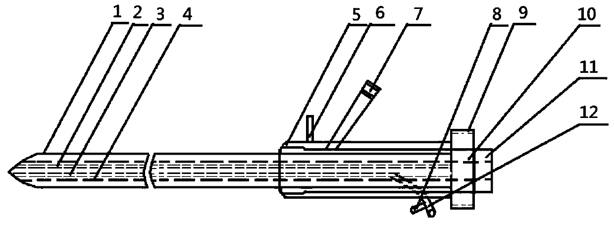
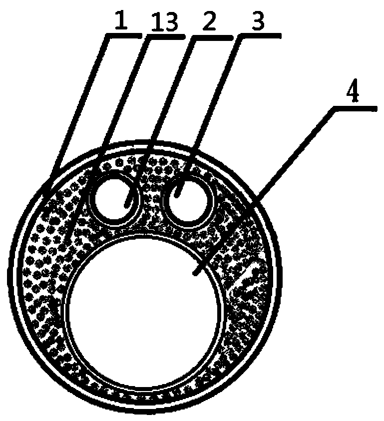
[0025] see Figure 1-2 , the present invention is an attractive ureteroscope, which consists of a ureteroscope front part 1, a camera channel 2, a water inlet and a holmium laser fiber channel 3, a water outlet and instrument channel 4, a ureteroscope middle and rear part 5, and a cold light source guide beam Connection port 6, camera eyepiece 7, water inlet pipe connector 8, water outlet and instrument channel joint thread 9, water outlet and instrument channel joint 10, water outlet and instrument channel sleeve 11, holmium laser fiber inlet 12, cold light source light guide fiber 13, Holmium laser fiber composition.

[0026] The front part 1 of the ureteroscope is connected with the back part 5 of the ureteroscope.

[0027] There is a cold light source light guide fiber interface 6 on one side of the front end of the rear part 5 of the ureteroscope; a camera eyepiece 7 is arranged behind the cold light guide light guide port 6; The laser fiber inlet 12 is obliquely connec...


PUM


Abstract

The invention relates to a urological surgical medical device, in particular to an attractive ureteroscope. An influent and holmium laser fiber channel is a cavity shared by the influent and holmium laser fibers during a surgery. An effluent and instrument channel penetrates the entire novel ureteroscope and is a cavity shared by the negative pressure effluent and instruments. An effluent and instrument channel joint is arranged on the rear portion in the ureteroscope, and the outer end of the effluent and instrument channel joint is provided with effluent and instrument channel joint threads.A sleeve of the effluent and instrument channel is arranged behind the effluent and instrument channel. The ureteroscope can accelerate the intraoperative drainage speed without affecting the surgical operation to form continuous water circulation in the surgical field, which can not only maintain the good definition of the visual field during the surgery, but also significantly improve the surgical lithotripsy efficiency. Further, the renal pelvis and ureteral pressure can be reduced to prevent postoperative bacteremia and urinary septicopyemia.

Description

technical field [0001] The invention relates to a medical device for urological surgery, in particular to a suctionable rigid ureteroscope. Background technique [0002] Ureteroscope is an endoscope used for minimally invasive surgery in urology. It is mainly used for the treatment of urinary system stones. During the operation, the ureteroscope is inserted into the bladder cavity through the urethra, and then enters the ureter cavity from the ureter opening. Exploration is carried out in the ureter, and the stones in the ureter are crushed using relevant lithotripsy instruments. [0003] The introduction of ultrasonic lithotripsy was a major advance. The early ultrasound probe was hollow, and the renal pelvis and ureter also sucked out the stones while crushing them. The diameter of the original probe is 8Fr, which cannot enter the working cavity of the ureteroscope. In this way, first use the ureteroscope to see the location of the stones, then take out the mirror and r...

Claims


Application Information

Patent Timeline
Patent Type & Authority Applications(China)
IPC IPC(8): A61B18/26A61B1/00A61B1/307A61B1/04A61B1/07A61B1/015
Inventor 范地福周毅
Owner 赣州市人民医院
Why Eureka
Try Eureka