Application of isoflavone in preparation of drug for treating fundus maculopathy
A technology for macular degeneration and isoflavones, applied in the field of biomedicine, can solve the problems of high cost and complicated operation, and achieve the effect of convenient medication, broad application prospects and significant curative effect
- Summary
- Abstract
- Description
- Claims
- Application Information
AI Technical Summary
Problems solved by technology
Method used
Image
Examples
Embodiment 1
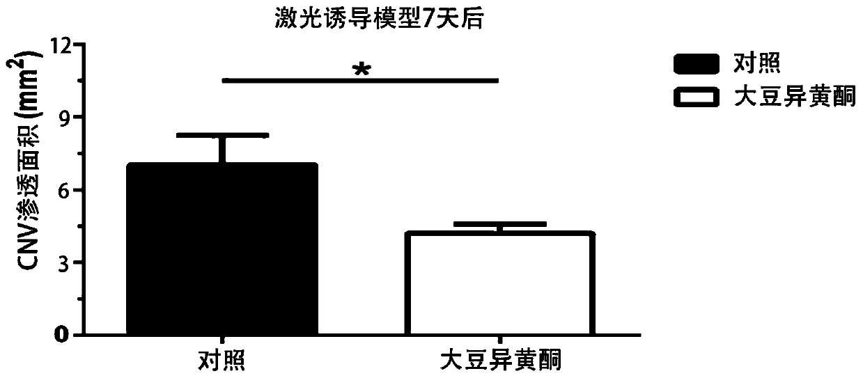
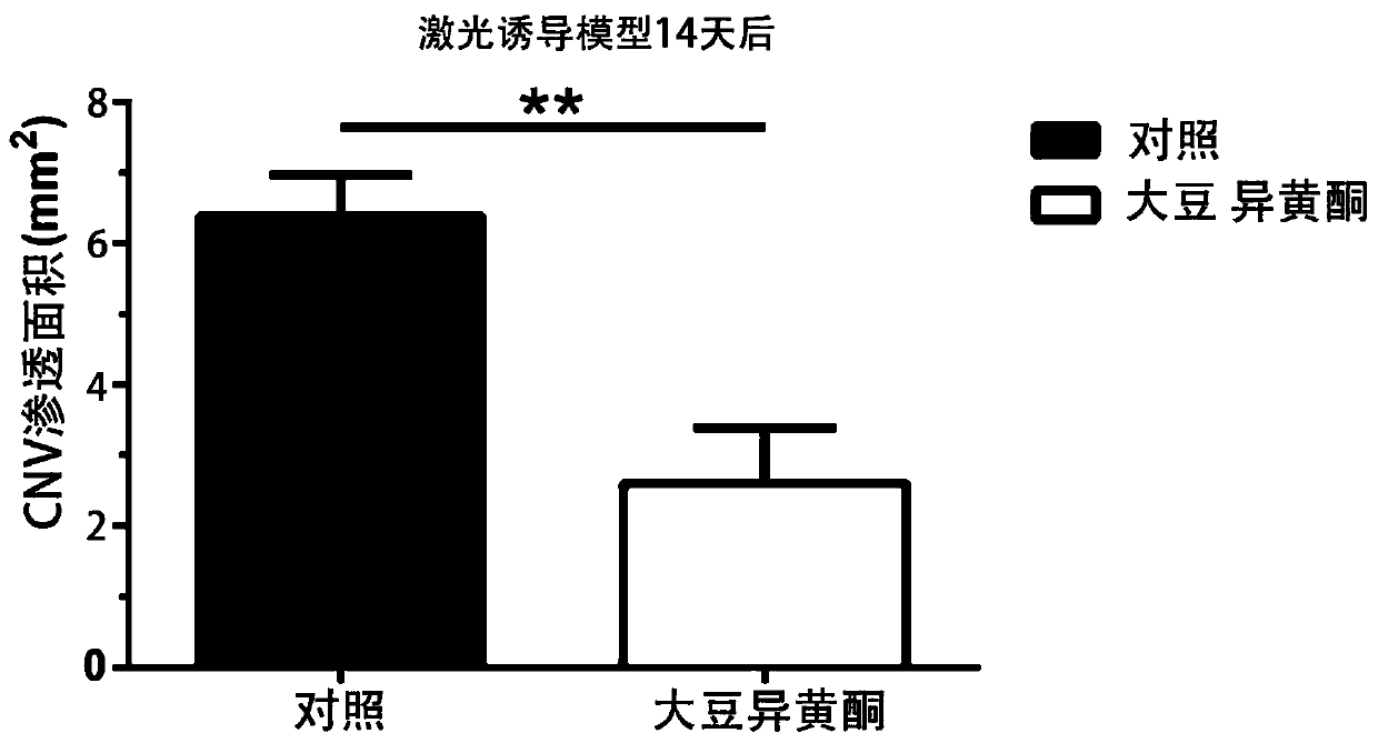
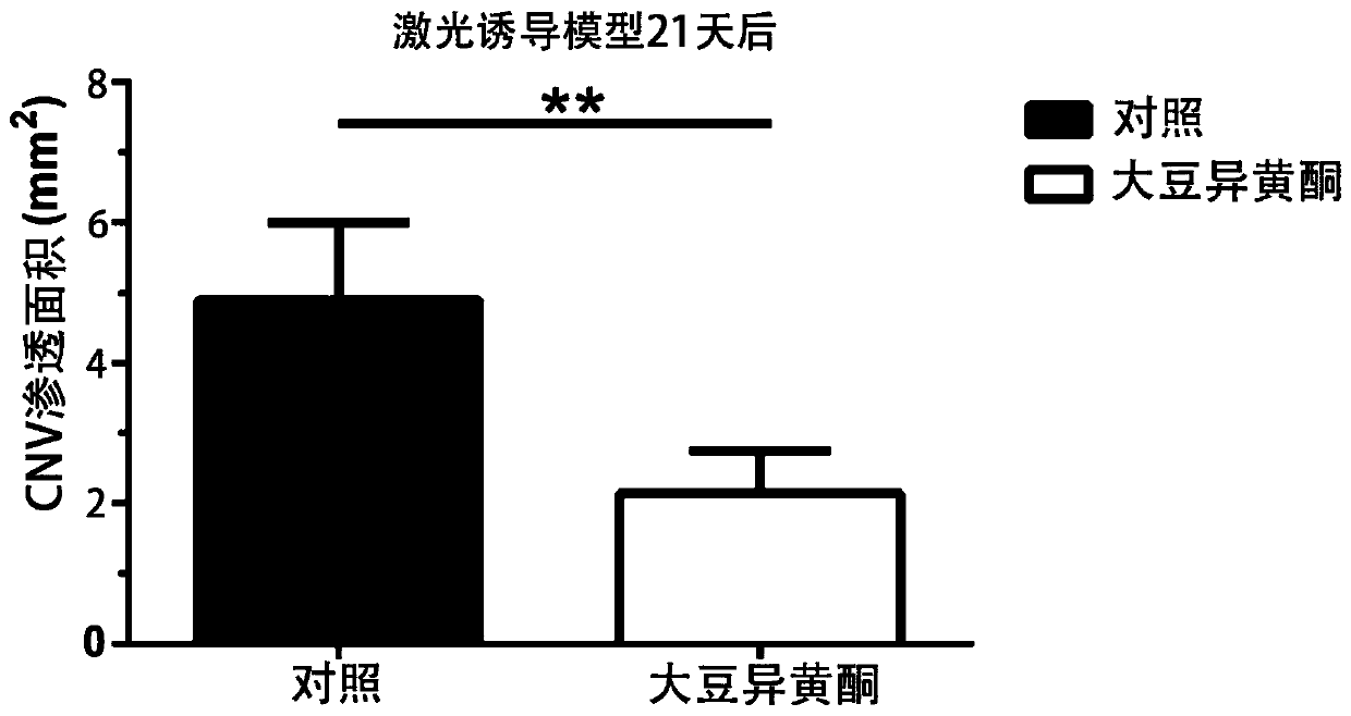
[0033] Choroidal neovascularization (CNV) model induced by 532nm argon laser was selected from C57 / B6 mice. (The laser point should avoid the main blood vessels and be 2.5 to 3.0 optic disc diameters away from the optic nerve head.) The mice with successful modeling were divided into two groups, each group n=10, and the experimental group was given 0.008% soybean isoflavone liquid ( 80 μg / mL), the control group was given the same volume of liquid matrix, and the volume of 50 μL per eye was locally administered to both eyes. On the 7th, 14th, and 21st day after the successful establishment of the mouse model, the choroidal vessels were measured by fluorescein angiography (FFA) and optical coherence tomography (OCT). The results of FFA combined with the results of OCT method were quantified and compared by t test. see results Figure 1 to Figure 4 .
[0034] Figure 1~3 The results showed that compared with the area of vascular infiltration after 7 days of laser-induced mo...
Embodiment 2
[0037] Choroidal neovascularization (CNV) model induced by 532nm argon laser was selected from C57 / B6 mice. (The laser point should avoid the main blood vessels and be 2.5 to 3.0 optic disc diameters away from the optic nerve head.) The mice with successful modeling were divided into two groups, each group n=10, and the experimental group was given 0.012% soybean isoflavone liquid ( 120 μg / mL), the control group was given the same volume of liquid matrix, and the volume of 50 μL per eye was locally administered to both eyes. On the 7th, 14th, and 21st day after the successful establishment of the mouse model, the choroidal vessels were measured by fluorescein angiography (FFA) and optical coherence tomography (OCT). The results of FFA combined with the results of OCT method were quantified and compared by t test.
[0038] Compared with the area of vascular infiltration area after 7 days of laser-induced model, the area of vascular infiltration in the isoflavone treatment ...
Embodiment 3
[0040]Select healthy colored rabbits, male or female, weighing 2 to 3 kg, and use an argon green laser with a wavelength of 514.5 nm (spot diameter of 50 μm, power of 0.7 W, and time of 0.1 s) to place a spot on the lateral medulla 2 to 3 diameters away from the optic disc. Intensively irradiate 20 points above and below the retina. The rabbits with successful modeling were divided into two groups, with n=10 in each group. The experimental group was given 0.005% soybean isoflavone liquid (50 μg / mL), and the control group was given the same volume of liquid base, with a volume of 50 μL per eye. Administer to both eyes. On the 7th, 14th, and 21st day after the successful establishment of the mouse model, the choroidal vessels were measured by fluorescein angiography (FFA) and optical coherence tomography (OCT). The results of FFA combined with the results of OCT method were quantified and compared by t test. see results Figure 5 to Figure 7 .
[0041] Figure 5-7 The resul...
PUM
 Abstract
Description
Claims
Application Information
 - R&D
- Intellectual Property
- Life Sciences
- Materials
- Tech Scout
- Unparalleled Data Quality
- Higher Quality Content
- 60% Fewer Hallucinations
Browse by: Latest US Patents, China's latest patents, Technical Efficacy Thesaurus, Application Domain, Technology Topic, Popular Technical Reports.
© 2025 PatSnap. All rights reserved.Legal|Privacy policy|Modern Slavery Act Transparency Statement|Sitemap|About US| Contact US: help@patsnap.com


