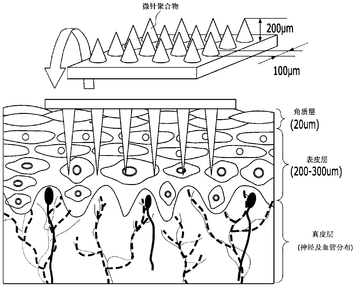
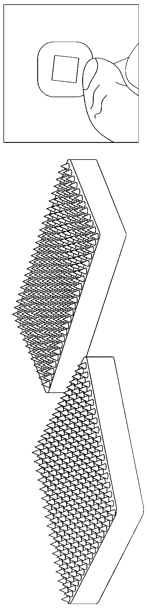
Method for fabricating microneedle-based diagnostic skin patch coated with aptamer and patch
A technology of aptamers and microneedles, applied in biochemical equipment and methods, diagnosis, microneedles, etc., can solve problems such as little effect
- Summary
- Abstract
- Description
- Claims
- Application Information
AI Technical Summary
Problems solved by technology
Method used
Image

Examples
Embodiment 1
[0038] Example 1. Reaction Example of Adhering Aptamers to the Surface of Microneedles
[0039] In order to avoid the uncertainty of diagnosis caused by non-specific binding to the protein of the microneedle, after the surface of the microneedle was treated with polyethylene glycol (PEG), the aptamer was adhered to the polyethylene glycol. end, thereby making the device.
[0040] A. Examples of polydimethylsiloxane microneedles
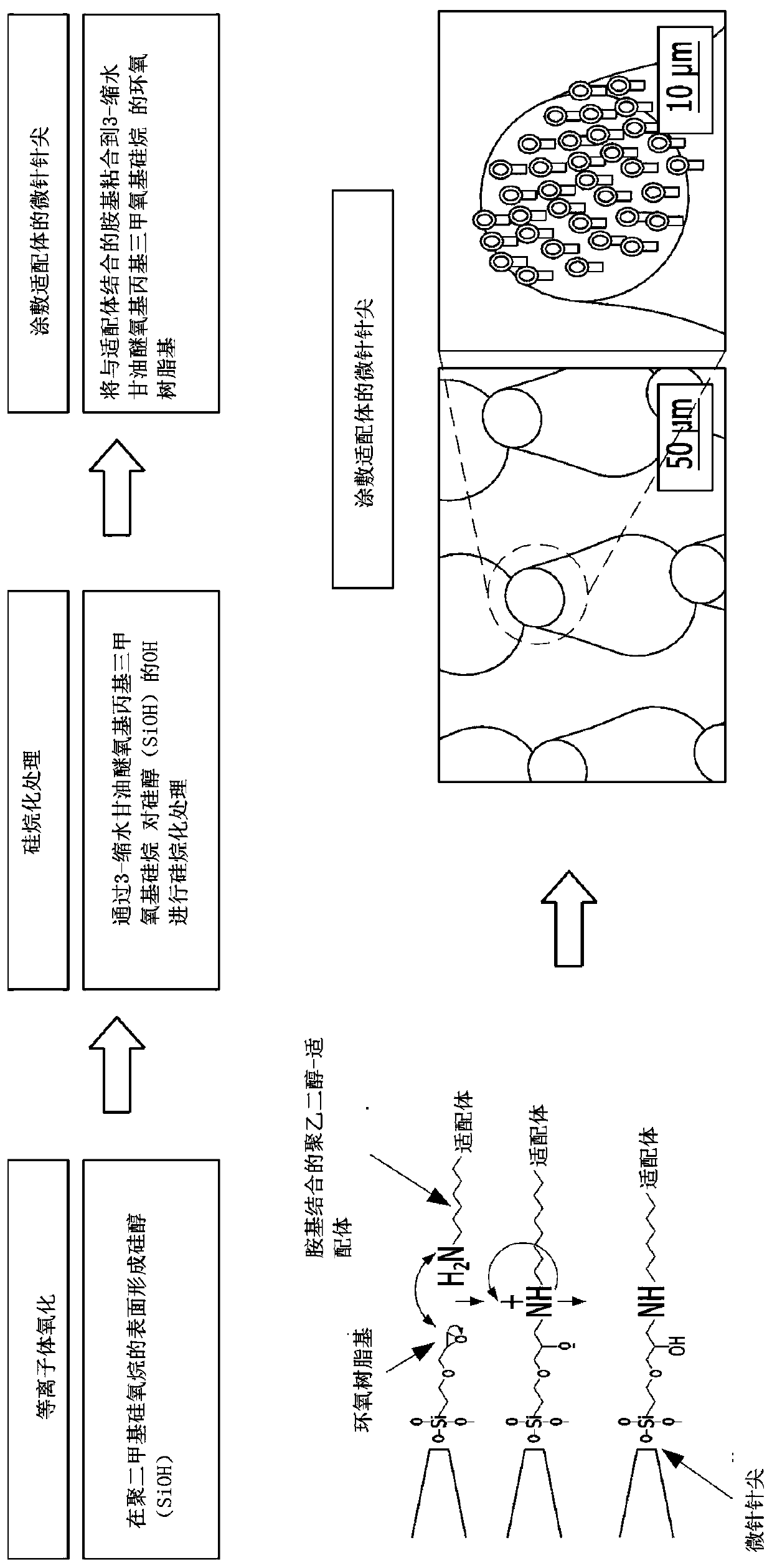
[0041] Silanol (SiOH) is formed on the surface of polydimethylsiloxane constituting the microneedles by plasma oxidation. After silylation treatment of the hydroxyl group by 3-glycidyl etheroxypropyl trimethoxysilane with epoxy resin group, the epoxy resin group and the amino group of polyethylene glycol combined with the aptamer ( Figure 4 ) combined, so that the aptamer-polyethylene glycol ( image 3 ).
[0042] B. Method for coating the surface of polycarbonate microneedles with an aptamer bound to one end of polyethylene glycol
[0043] The ...
PUM
 Abstract
Description
Claims
Application Information
 - R&D Engineer
- R&D Manager
- IP Professional
- Industry Leading Data Capabilities
- Powerful AI technology
- Patent DNA Extraction
Browse by: Latest US Patents, China's latest patents, Technical Efficacy Thesaurus, Application Domain, Technology Topic.
© 2024 PatSnap. All rights reserved.Legal|Privacy policy|Modern Slavery Act Transparency Statement|Sitemap