Method for optical imaging of living myocardial sheet

A technology of optical imaging and myocardium, applied in medical science, scientific instruments, fluorescence/phosphorescence, etc.

Pending Publication Date: 2019-08-16
SOUTHWEST MEDICAL UNIVERISTY
View PDF0 Cites 2 Cited by
  • Summary
  • Abstract
  • Description
  • Claims
  • Application Information

AI Technical Summary

Problems solved by technology

However, the combination of optogenetics, optical mapping and living myocardium slices has not yet been realized to achieve non-invasive ...

Method used


Image

  • Method for optical imaging of living myocardial sheet
  • Method for optical imaging of living myocardial sheet
  • Method for optical imaging of living myocardial sheet

Examples


Embodiment 1

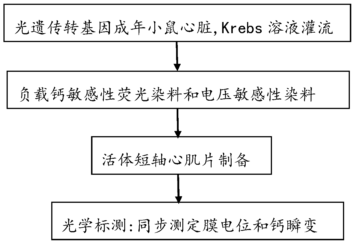
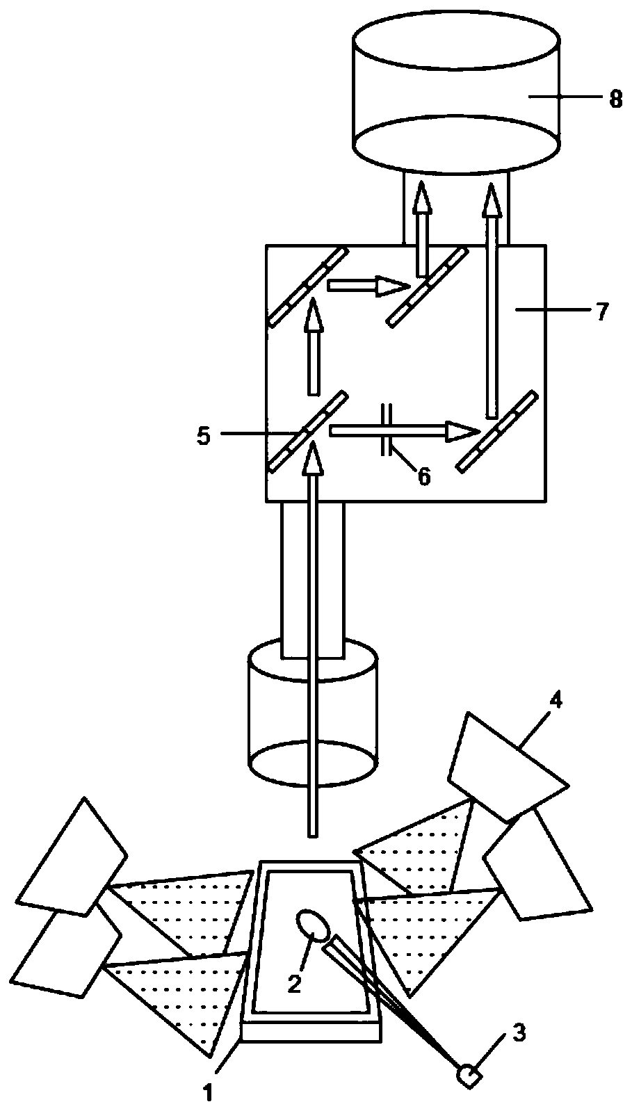
[0106] The method for optical imaging of living myocardial slices in this embodiment, such as figure 1 shown, including the following steps:

[0107] Step 1: Perfusion of isolated hearts

[0108] Step 1.1: Take the optogenetic transgenic adult mouse and anesthetize it.

[0109] The optogenetic transgenic adult mice were composed of Pnmt-Cre transgenic mice and B6.Cg-Gt(ROSA)26Sor tm27.1 (CAG-COP4H134R / tdTomato)Hze / J strain mouse cross. The DNA between the loxP sites is cut by Cre recombinase, and the nick is reconnected under the action of DNA ligase. After recombination, hChR2(H134R)-tdTomato is connected behind the Pnmt promoter, that is, Pnmt and hChR2(H134R)- are simultaneously expressed. td Tomato. The above-mentioned optogenetic transgenic adult mice were purchased from Oxford University. The Pnmt-Cre transgenic mouse inserts Cre recombinase on the Pnmt gene promoter, and the B6.Cg-Gt(ROSA)26Sor tm27.1(CAG-COP4H134R / tdTomato)Hze / J strain mice have CAG-loxP-STOP-...

Embodiment 2

[0128] The method for optical imaging of living myocardial slices in this embodiment, such as figure 1 shown, including the following steps:

[0129] Step 1: Perfusion of isolated hearts

[0130] Step 1.1: Take the optogenetic transgenic adult mouse and anesthetize it.

[0131] The optogenetic transgenic adult mice were composed of Pnmt-Cre transgenic mice and B6.Cg-Gt(ROSA)26Sor tm27.1 (CAG-COP4H134R / tdTomato)Hze / J strain mouse cross. The DNA between the loxP sites is cut by Cre recombinase, and the nick is reconnected under the action of DNA ligase. After recombination, hChR2(H134R)-tdTomato is connected behind the Pnmt promoter, that is, Pnmt and hChR2(H134R)- are simultaneously expressed. td Tomato. The above-mentioned optogenetic transgenic adult mice were purchased from Oxford University. The Pnmt-Cre transgenic mouse inserts Cre recombinase on the Pnmt gene promoter, and the B6.Cg-Gt(ROSA)26Sor tm27.1(CAG-COP4H134R / tdTomato)Hze / J strain mice have CAG-loxP-STOP-...

Embodiment 3

[0150] The method for optical imaging of living myocardial slices in this embodiment, such as figure 1 shown, including the following steps:

[0151] Step 1: Perfusion of isolated hearts

[0152] Step 1.1: Take the optogenetic transgenic adult mouse and anesthetize it.

[0153] The optogenetic transgenic adult mice were composed of Pnmt-Cre transgenic mice and B6.Cg-Gt(ROSA)26Sor tm27.1 (CAG-COP4H134R / tdTomato)Hze / J strain mouse cross. The DNA between the loxP sites is cut by Cre recombinase, and the nick is reconnected under the action of DNA ligase. After recombination, hChR2(H134R)-tdTomato is connected behind the Pnmt promoter, that is, Pnmt and hChR2(H134R)- are simultaneously expressed. td Tomato. The above-mentioned optogenetic transgenic adult mice were purchased from Oxford University. The Pnmt-Cre transgenic mouse inserts Cre recombinase on the Pnmt gene promoter, and the B6.Cg-Gt(ROSA)26Sor tm27.1(CAG-COP4H134R / tdTomato)Hze / J strain mice have CAG-loxP-STOP-...


PUM

PropertyMeasurementUnit
Thicknessaaaaaaaaaa
Thicknessaaaaaaaaaa
Thicknessaaaaaaaaaa

Abstract

The invention discloses a method for optical imaging of a living myocardial sheet, and belongs to the technical field of optical imaging. The method for optical imaging of a living myocardial sheet comprises the following steps of step 1: perfusion of an isolated heart; step 2: loading of a calcium-sensitive fluorescent dye and a voltage-sensitive fluorescent dye; step 3: preparation of a living short-axis myocardial sheet; and step 4: optical mapping. The method combines optogenetic technique, optical mapping and living myocardial sheet technology, performs the short-axis sectioning of the heart of a photogenetical transgenic mouse, and adopts an ultra-high-rate camera for optical mapping for detecting and analyzing the conduction characteristics of myocardial membrane potential and calcium transients after blue light stimulation, thereby opening up a new research method for the physiology and pathology of the heart.

Description

technical field [0001] The invention relates to a method for optical imaging of living myocardial slices, belonging to the technical field of optical imaging. Background technique [0002] Basic research on the heart mainly uses traditional biological models ranging from isolated cardiomyocytes to complex multicellular tissues, including isolated hearts and intact tissue blocks (such as isolated myocardial strips and perfused myocardial blocks). However, isolated cardiomyocytes disrupt the connection between cardiomyocytes and the extracellular matrix during the enzymatic separation process, which seriously affects the electrophysiological function of cardiomyocytes; multicellular tissues have problems related to in vitro perfusion and tissue ischemia. [0003] Optogenetics is the use of light to activate light-sensitive ion channels and opsins to depolarize or hyperpolarize cells and control the "on" and "off" of excitable cells. This emerging technology is grounded in neu...

Claims


Application Information

Patent Timeline
IPC IPC(8): G01N21/64G01N1/30G01N1/06G01N1/28A61D1/00
CPCA61D1/00G01N1/06G01N1/286G01N1/30G01N21/64G01N2001/2873
Inventor 雷鸣文强欧贤红
Owner SOUTHWEST MEDICAL UNIVERISTY
Why Eureka
Try Eureka