Combined formula kit for analyzing phenotype and function of CD1c+ dendritic cell subsets and application thereof
A dendritic cell and kit technology, which is applied in the biological field, can solve the problem of not being able to study CD1c+DC subgroups, etc., and achieve the effect of rapid one-step determination, long time consumption, and saving a lot of time.
- Summary
- Abstract
- Description
- Claims
- Application Information
AI Technical Summary
Problems solved by technology
Method used
Image


Examples
Embodiment 1
[0054] Example 1 Pretreatment of Non-small Cell Lung Cancer Patient / Healthy People's Peripheral Blood
[0055] The preprocessing steps are as follows:
[0056] (1) Take 1 drop (10-100 μl) of venous peripheral blood from patients with non-small cell lung cancer and healthy adults, and perform anticoagulant treatment;
[0057] (2) Mix peripheral whole blood with 2 ml of 1× red blood cell lysate (Biolegend), rotate and oscillate for 10 seconds, and let stand at room temperature in the dark for 15 minutes;
[0058] (3) centrifuge (350g * 5 minutes) with centrifuge, pour over the supernatant, then the precipitated cells are suspended in 2 milliliters of cell staining fluid (containing the PBS solution of 2.5% fetal bovine serum);
[0059] (4) Leukocyte-stimulating factor (BD) was added at a concentration ratio of 0.1%, and the cells were incubated at a constant temperature of 37 degrees for 6 hours for later use.
Embodiment 2
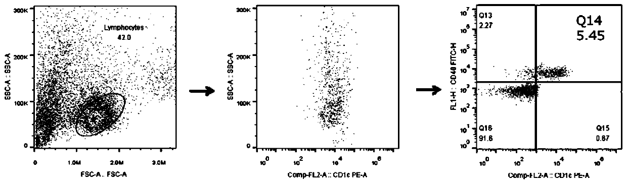
[0060] Example 2 Analysis of the degree of development and differentiation of CD1c+ dendritic cell subsets in peripheral blood of non-small cell lung cancer patients / healthy people
[0061] The spare blood cells were centrifuged (350 g) for 5 minutes, the supernatant was decanted, and the cells were suspended in 100 μl of cell staining solution. Then add 2 μl of anti-human CD1c and CD40 antibodies (Biolegend), incubate at room temperature for 30 minutes, then add 2 ml of cell staining solution, and repeat centrifugation twice at 350 g for 5 minutes each time. After decanting the supernatant, fix the cells with 2 ml of 2% formalin solution, and incubate at room temperature in the dark for 20 minutes for later use.
Embodiment 3
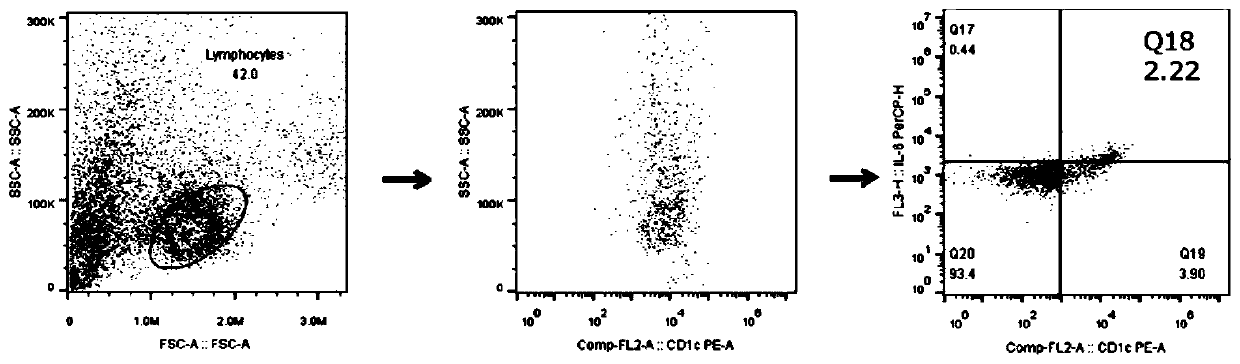
[0062] Example 3 Functional Analysis of Peripheral Blood CD1c+ Dendritic Cell Subsets of Non-small Cell Lung Cancer Patients / Healthy People
[0063] (1) Suspend the fixed spare cells in 2 ml of cell penetration solution (Biolegend), then repeat centrifugation (350g) for 10 minutes twice;
[0064] (2) Resuspend the pelleted cells after centrifugation in 100 μl of cell permeation buffer, then add 2 μl of IL-6 (Biolegend) and IL-10 antibody (BD), and incubate at room temperature for 30 minutes in the dark;
[0065] (3) The incubated cells were suspended in 2 ml of cell penetration solution, and then centrifuged (350g) for 5 minutes twice;
[0066] (4) Finally, decant the supernatant, resuspend the pelleted cells in 0.5 ml of cell staining solution, and analyze and test with a flow cytometer.
PUM
| Property | Measurement | Unit |
|---|---|---|
| Volume | aaaaa | aaaaa |
Abstract
Description
Claims
Application Information
 - R&D Engineer
- R&D Manager
- IP Professional
- Industry Leading Data Capabilities
- Powerful AI technology
- Patent DNA Extraction
Browse by: Latest US Patents, China's latest patents, Technical Efficacy Thesaurus, Application Domain, Technology Topic.
© 2024 PatSnap. All rights reserved.Legal|Privacy policy|Modern Slavery Act Transparency Statement|Sitemap