Image scanning microscopic imaging method and device based on double-microlens array

A micro-lens array and image scanning technology, applied in microscopes, measuring devices, instruments, etc., can solve the problems of complex reconstruction algorithms, slow imaging speed of image scanning microscopy imaging technology, and reduction of reconstruction time, so as to improve scanning speed, improve Data collection efficiency, the effect of reducing reconstruction time

Inactive Publication Date: 2020-04-07
HARBIN INST OF TECH
View PDF5 Cites 2 Cited by
  • Summary
  • Abstract
  • Description
  • Claims
  • Application Information

AI Technical Summary

Problems solved by technology

[0006] In order to solve the problems of slow imaging speed and complex subsequent reconstruction algorithm of image scanning microscopic imaging technology, the present invention designs an image scanning microscopic imaging method and device based on a double microlens array. The excitation beam is generated by the first micro

Method used


Image

  • Image scanning microscopic imaging method and device based on double-microlens array

Examples


Embodiment Construction

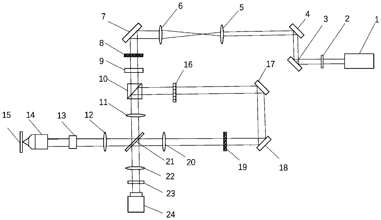
[0025] Embodiments of the present invention will be described in detail below in conjunction with the accompanying drawings

[0026] The image scanning microscopic imaging method and device based on the double microlens array of this embodiment includes a laser 1, and a half-wave plate 2 is placed in front of the laser 1 to realize the intensity control of the laser, and a first lens 5 is arranged on the direct optical path of the laser The beam expander that forms with the second lens 6, the light after beam expansion is directed to the first microlens array 8, and this first microlens array focal length is 2mm, and diameter is 25mm, is provided with compensating plate 9 thereafter, in order to eliminate Astigmatism of a focused beam. The multi-focus illumination is collimated through the first scanning lens 11 and the second scanning lens 12, which constitute a 4f system. The excitation beam of the sample 15 plane is imaged through the tubular lens 13 and the objective lens...


PUM


Abstract

The invention relates to an image scanning microscopic imaging method and device based on a double-microlens array, and belongs to the technical field of optical microscopic measurement. According tothe invention, an array type multi-focus generated by the micro-lens array is used for illuminating a sample; the imaging speed is improved, the influence of out-of-focus fluorescence is eliminated through the pinhole array, each focusing light beam is scaled by 1/2 through the second micro lens array, all-optical implementation of subsequent digital information processing of traditional image scanning microscopy is achieved, it is guaranteed that the resolution ratio is improved by two times, and meanwhile the problem that a traditional image scanning microscopy system is low in imaging speedis solved. The laser beam reaches the first micro-lens array after being expanded to generate an array type illumination beam, and after the array type illumination beam is emitted to the surface ofa sample, the objective lens collects multi-focus fluorescence from the sample and emits the multi-focus fluorescence to the pinhole array to eliminate the influence of out-of-focus fluorescence. Thesecond micro-lens array locally shrinks each focus point, meanwhile, the original focusing direction of the light beam is kept, and the zoomed fluorescence focus is imaged to the sCMOS camera under the deflection effect of the scanning galvanometer.

Description

technical field [0001] An image scanning microscopic imaging method and device based on a double microlens array belongs to the field of optical microscopic measurement, and mainly relates to a method and a device for non-contact rapid imaging of fine structures of biological samples based on microstructured optical elements. Background technique [0002] Modern fluorescence microscopy combines imaging speed, molecular specificity, and contrast to visualize the cellular imaging process, but diffraction limits the resolution of wide-field microfluorescence microscopy, while super-resolution imaging breaks through the traditional diffraction limit, but in There is a decrease in speed, imaging time and field of view. For example, single-molecule imaging counting and stimulated emission depletion microscopy enable cells with a spatial resolution of less than 100 nm, but imaging speeds are limited to 0.01-1 Hz. [0003] Compared with single-molecule imaging counting and stimulat...

Claims


Application Information

Patent Timeline
IPC IPC(8): G02B21/00G02B21/36G02B21/06G01N21/64
CPCG01N21/6458G02B21/0032G02B21/0076G02B21/06G02B21/361
Inventor 王伟波张宝元吴必伟谭久彬
Owner HARBIN INST OF TECH
Why Eureka
Try Eureka