Calprotectin monoclonal antibody and application thereof
A monoclonal antibody and calprotectin technology, applied in the field of immunology, can solve the problems of cumbersome operation, time-consuming detection, inconvenient movement of detection equipment, etc., and achieve the effect of high detection sensitivity and good cross-linking effect
- Summary
- Abstract
- Description
- Claims
- Application Information
AI Technical Summary
Problems solved by technology
Method used
Image
Examples
Embodiment 1
[0045] An embodiment of the present invention provides a calprotectin monoclonal antibody, which comprises VHCDR1-3 of the heavy chain variable region and VLCDR1-3 of the light chain variable region;
[0046] VHCDR1: GGATTCACTAGTAGCTATGCC
[0047] VHCDR2: ATTAGTAGTTATGGTAGCACC
[0048] VHCDR3: GCAACCTACAGGTACGAAGGGTTTGCTTAC
[0049] VLCDR1: ACTGGGGCTGTTACAACTAGTAACTAT
[0050] VLCDR2: GGTACCAAC
[0051] VLCDR3: GCTCTATGGTACAGCAACCTTTGGGTG.
[0052] Wherein, the amino acid sequences of VHCDR1, VHCDR2 and VHCDR3 are respectively shown in SEQ ID NOs: 1-3:
[0053] SEQ ID NO: 1Gly-Phe-Thr-Ser-Ser-Tyr-Ala
[0054]SEQ ID NO: 2Ile-Ser-Ser-Tyr-Gly-Ser-Thr
[0055] SEQ ID NO: 3Ala-Thr-Tyr-Arg-Tyr-Glu-Gly-Phe-Ala-Tyr.
[0056] Wherein, the amino acid sequences of VLCDR1, VLCDR2 and VLCDR3 are respectively shown in SEQ ID NOs: 4-6:
[0057] SEQ ID NO: 4Thr-Gly-Ala-Val-Thr-Thr-Ser-Asn-Tyr
[0058] SEQ ID NO: 5Gly-Thr-Asn
[0059] SEQ ID NO: 6Ala-Leu-Trp-Tyr-Ser-Asn-Leu-Trp-Val. ...
Embodiment 2
[0061] The present invention further discloses a preparation method of the above-mentioned mouse anti-human calprotectin monoclonal antibody, and the preparation method comprises the following steps:
[0062] The calprotectin monoclonal antibody is secreted by a hybridoma cell line; the preparation method of the hybridoma cell line comprises the following steps:
[0063] 1) Using natural calprotectin extracted from neutrophils as immune antigen;
[0064] 2) immunizing mice with the natural calprotectin prepared in 1);
[0065] 3) performing cell fusion of myeloma cell suspension and spleen B lymphocytes of immunized mice;
[0066] 4) culturing the fused cells, measuring the titer of the hybridomas, screening with natural calprotectin polymers, eliminating clones that react with protein monomers, and obtaining the hybridoma cell lines;
[0067] In order to overcome the problems of instability and degeneration of the hybridoma cell line, the hybridoma cell line is extracted fr...
Embodiment 3
[0095] The embodiment of the present invention discloses the application of the above-mentioned calprotectin monoclonal antibody in the immune detection tool of inflammatory bowel disease;
[0096] Immunological detection tools include inflammatory bowel disease detection kits.
[0097] (1) Calprotectin Immunoturbidimetric Quantitative Detection Kit
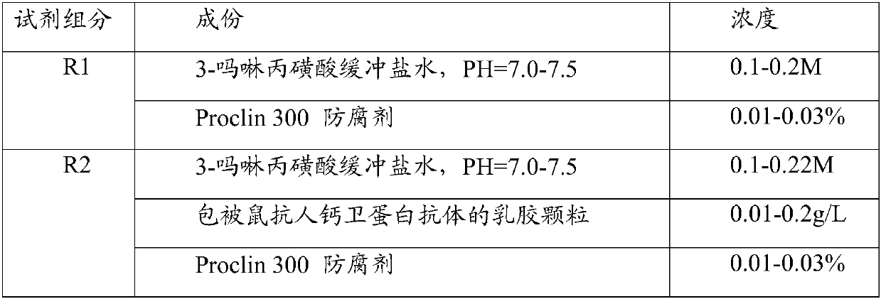
[0098] In this example, the calprotectin monoclonal antibody obtained in Example 1 is labeled with latex microspheres, the particle size is 200-300nm, and the microsphere concentration is 0.2-1.5% prepared with 3-morpholine propanesulfonic acid buffer solution, buffered The agent contains antibody stabilizers, preservatives and other components.
[0099] The preparation and operation of the calprotectin in vitro diagnostic kit are as follows:
[0100] 1. Preparation of R1 reagent:
[0101] 3-Morpholine propanesulfonic acid buffered saline solution: 0.2-1.0M, pH7.0-7.5 3-morpholine propanesulfonic acid buffer
[0102] 2. Prepa...
PUM
| Property | Measurement | Unit |
|---|---|---|
| Particle size | aaaaa | aaaaa |
Abstract
Description
Claims
Application Information
 - R&D
- Intellectual Property
- Life Sciences
- Materials
- Tech Scout
- Unparalleled Data Quality
- Higher Quality Content
- 60% Fewer Hallucinations
Browse by: Latest US Patents, China's latest patents, Technical Efficacy Thesaurus, Application Domain, Technology Topic, Popular Technical Reports.
© 2025 PatSnap. All rights reserved.Legal|Privacy policy|Modern Slavery Act Transparency Statement|Sitemap|About US| Contact US: help@patsnap.com


