A facile protein purification method for localizing recombinant proteins to the cell surface

A recombinant protein and cell surface technology, applied in the field of simple purification of protein, to save time and cost, and simple operation

Active Publication Date: 2021-02-23
BIOINTRON BIOLOGICAL INC
View PDF3 Cites 0 Cited by
  • Summary
  • Abstract
  • Description
  • Claims
  • Application Information

AI Technical Summary

Problems solved by technology

[0006] Aiming at the defect in the prior art that recombinant proteins require complex purification steps to obtain relatively pure proteins after they are expressed in cells, the technical problem to be solved in this application is to provide a method for quickly obtaining high-purity proteins without lysing cells and purifying through chromatographic columns. The method of the target protein, the method is to fuse and express the gene encoding the cell membrane anchor protein (signal peptide), the chemical method cleavage site and the target protein, so that the fusion protein is positioned on the surface of the cell

Method used


Image

  • A facile protein purification method for localizing recombinant proteins to the cell surface
  • A facile protein purification method for localizing recombinant proteins to the cell surface
  • A facile protein purification method for localizing recombinant proteins to the cell surface

Examples


Embodiment 1

[0044] Embodiment 1: Construction and expression of Lpp-OmpA-(SNAC-tag)-GB1 fusion protein and Lpp-OmpA-(SNAC-tag)-SFGFP fusion protein

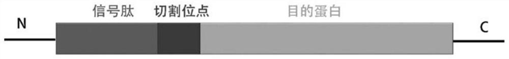
[0045] The mode of the fusion protein used in this embodiment is as follows figure 1 As shown, the code Lpp(1-9)-OmpA(45-159)-(SNAC-tag)-GB1 (sequence shown in SEQ ID NO.1) obtained by gene synthesis (Sangon Bioengineering Co., Ltd.) Shown) the pet30a plasmid was transformed into E.coli BL21(DE3), the expression was induced by 0.5mM IPTG at 30°C for 5h, and the bacteria were harvested by a high-speed centrifuge. and stored at -80°C until use. The pet30a plasmid encoding Lpp(1-9)-OmpA(45-159)-(SNAC-tag)-SFGFP (sequence shown in SEQ ID NO.2) was obtained by total gene synthesis (Sangon Bioengineering Co., Ltd.) Express in the same way and collect bacteria.

Embodiment 2

[0046] Embodiment 2: Construction, expression and purification of 6His-GB1 fusion protein and 6His-SFGFP fusion protein

[0047] The pet28a plasmid encoding 6His-GB1 (sequence shown in SEQ ID NO.3) obtained by gene synthesis (Sangon Bioengineering Co., Ltd.) was transformed into E.coli BL21 (DE3), using 0.5mM IPTG in The expression was induced at 30°C for 5 hours, and the bacteria were harvested using a high-speed centrifuge. and stored at -80°C until use. The pet28a plasmid encoding 6His-SFGFP (sequence shown in SEQ ID NO.4) was obtained by total gene synthesis (Sangon Bioengineering Co., Ltd.) and obtained and expressed in the same manner, and the bacteria were harvested.

Embodiment 3

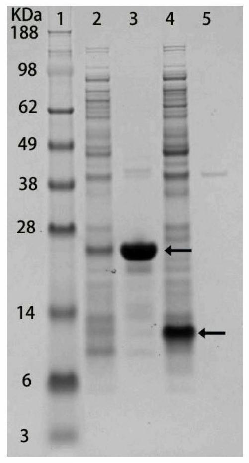
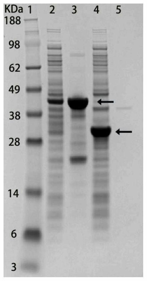
[0048] Example 3: Identification of cell membrane and cytoplasmic components of bacteria expressing fusion protein

[0049] In order to characterize whether the signal peptide can localize the target protein to the cell membrane, it is necessary to identify the cell membrane and cytoplasmic components of the bacteria expressing the recombinant protein by SDS-PAGE gel. Bacteria were resuspended in 10 mM PBS (pH 7.4) and lysed using sonication. The supernatant and the precipitate were separated by high-speed centrifugation at 12000 rpm, and the precipitate was eluted twice with TDSET buffer (1% Triton X-100, 0.2% sodium deoxycholate, 0.1% SDS, 10 mM tetrasodium EDTA, and 10 mM Tris / HCl). Pellets and supernatants were identified using SDS-PAGE. figure 2 The 2nd lane is the supernatant of bacteria expressing Lpp-OmpA-(SNAC-tag)-GB1, the 3rd lane is the cell membrane components of Lpp-OmpA-(SNAC-tag)-GB1 bacteria, and the 4th lane is expressing 6His- The supernatant of GB1 bacte...


PUM


Abstract

This application discloses a simple protein purification method for positioning recombinant proteins on the cell surface, which is characterized in that, by constructing a gene encoding a signal peptide, a chemical cleavage site, and a target protein into an expression vector, the target protein is expressed Locate to the surface of the cells; add the buffer containing the cleavage reagent to the collected cells to resuspend, and the supernatant after centrifugation is the target protein solution. The simple protein purification method of the present application utilizes the localization function of the signal peptide, so that the recombinant protein is secreted and localized to the surface of the cell after being expressed in the cell. And taking advantage of the chemical cleavage site in the middle of the recombinant protein, you only need to add a cleavage reagent to the buffer used to resuspend the cells, and centrifuge to obtain a high-purity target protein very easily. It solves the cumbersome steps of lysing cells and using chromatographic columns of various properties to obtain the target protein in the prior art for protein purification, and the operation is simple, which greatly saves time and cost.

Description

technical field [0001] The application belongs to the field of protein separation and purification, and relates to a simple method for purifying protein by positioning recombinant protein on the cell surface. By constructing the fusion gene encoding the signal peptide, the chemical cleavage site and the target protein into the expression vector, the target protein can be localized to the outer membrane of the cell after expression. The recombinant protein expressed by this method only needs to be resuspended in the buffer containing the cleavage reagent in the collected cells, without lysing the cells and purifying the chromatographic column, and can obtain the target protein with high purity and high yield in one step after centrifugation. Background technique [0002] The expression systems of recombinant proteins can be divided into four types: prokaryotic, mammalian, yeast and insect expression systems. The expression sites of recombinant proteins in cells can be divide...

Claims


Application Information

Patent Timeline
Patent Type & Authority Patents(China)
IPC IPC(8): C07K19/00C12N15/70C12P21/00C07K1/14
CPCC07K14/00C07K2319/02C07K2319/21C12N15/70C12P21/00
Inventor 黄善青沙万星刘天意魏天彪
Owner BIOINTRON BIOLOGICAL INC
Why Eureka
Try Eureka