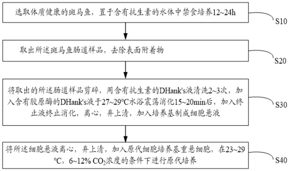
Method for separating and primary culture of zebrafish intestinal mucosal epithelial cells
A primary culture, intestinal mucosa technology, applied in cell dissociation methods, cell culture active agents, gastrointestinal cells, etc.
- Summary
- Abstract
- Description
- Claims
- Application Information
AI Technical Summary
Problems solved by technology
Method used
Image
Examples
Embodiment 1
[0052] Select healthy zebrafish and place them in water containing 80,000 U / L penicillin and 80,000 U / L gentamicin for fasting culture for 12 hours. Take the fasted zebrafish and wash them with ultrapure water Tap the zebrafish brain twice to kill it, and then quickly soak it in 70% ethanol for 20-30s. Open the zebrafish abdomen on an ultra-clean bench, take out the zebrafish foregut, and remove the surface attachments.
[0053] Cut the removed zebrafish foregut into pieces of 1-2 mm 3 Tissue blocks were washed 2-3 times with D-Hank's solution containing 80,000 U / L penicillin and 80,000 U / L gentamycin, and then added with 0.2 g / L collagenase Ⅰ and 0.2 g / L collagenase Ⅳ D-Hank's solution was shaken and digested in a water bath at 28°C for 15 minutes, then stop solution (5% FBS+95% DMEM medium) was added to stop the digestion, centrifuged at 400r / min for 5 minutes, the supernatant was discarded, and DMEM medium was added to make a cell suspension.
[0054] Centrifuge the above ...
Embodiment 2
[0056] Select healthy zebrafish and place them in water containing 80,000 U / L penicillin and 80,000 U / L gentamicin for fasting culture for 24 hours. Take the fasted zebrafish and wash them with ultrapure water. Tap the zebrafish brain twice to kill it, and then quickly soak it in 70% ethanol for 20-30s. Open the zebrafish abdomen on an ultra-clean bench, take out the zebrafish foregut, and remove the surface attachments.
[0057] Cut the removed zebrafish foregut into pieces of 1-2 mm 3 The tissue block was washed 2 to 3 times with D-Hank's solution containing 80,000 U / L penicillin and 80,000 U / L gentamicin, and then added with 0.25 g / L collagenase Ⅰ and 0.25 g / L collagenase Ⅳ D-Hank's solution was shaken and digested in a water bath at 27°C for 18 minutes, then stop solution (5% FBS+95% DMEM medium) was added to stop the digestion, centrifuged at 350r / min for 8 minutes, the supernatant was discarded, and DMEM medium was added to make a cell suspension.
[0058] The above-men...
Embodiment 3
[0060] Select healthy zebrafish and place them in water containing 80,000 U / L penicillin and 80,000 U / L gentamicin for fasting culture for 18 hours. Take the fasted zebrafish and wash them with ultrapure water Tap the zebrafish brain three times to kill it, and then quickly soak it in 70% ethanol for 20-30 seconds. Open the zebrafish abdomen on an ultra-clean bench, take out the zebrafish foregut, and remove surface attachments.
[0061] Cut the removed zebrafish foregut into pieces of 1-2 mm 3 Tissue blocks were washed 2-3 times with D-Hank's solution containing 80,000 U / L penicillin and 80,000 U / L gentamicin, and then added with 0.15 g / L collagenase Ⅰ and 0.15 g / L collagenase Ⅳ D-Hank's solution was shaken and digested in a water bath at 29°C for 20 minutes, then stop solution (5% FBS+95% DMEM medium) was added to stop the digestion, centrifuged at 500r / min for 4 minutes, the supernatant was discarded, and DMEM medium was added to make a cell suspension.
[0062]The above c...
PUM
 Abstract
Description
Claims
Application Information
 - R&D
- Intellectual Property
- Life Sciences
- Materials
- Tech Scout
- Unparalleled Data Quality
- Higher Quality Content
- 60% Fewer Hallucinations
Browse by: Latest US Patents, China's latest patents, Technical Efficacy Thesaurus, Application Domain, Technology Topic, Popular Technical Reports.
© 2025 PatSnap. All rights reserved.Legal|Privacy policy|Modern Slavery Act Transparency Statement|Sitemap|About US| Contact US: help@patsnap.com

