
Methods and uses for promoting skeletal muscle development

A skeletal muscle and gene technology, applied in the field of promoting skeletal muscle development, can solve problems such as unclear regulatory mechanism, achieve wide applicability, promote skeletal muscle development, and promote development.

Active Publication Date: 2021-12-21
SOUTH CHINA AGRI UNIV
View PDF6 Cites 0 Cited by
  • Summary
  • Abstract
  • Description
  • Claims
  • Application Information

AI Technical Summary

Problems solved by technology

In recent years, the regulation of H3K27me3 modification on muscle development has attracted much attention. A large number of studies have shown that H3K27me3 plays an important role in regulating the development of skeletal muscle. However, the specific regulatory mechanism is still unclear. few

Method used


Image

  • Methods and uses for promoting skeletal muscle development
  • Methods and uses for promoting skeletal muscle development
  • Methods and uses for promoting skeletal muscle development

Examples


Embodiment 1

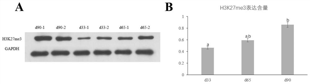
[0029] Example 1 Detection of H3K27me3 expression level in pig skeletal muscle samples in 3 different periods

[0030] A total of 6 tissue samples of the longissimus dorsi muscle samples of Duroc pig embryos at 33 days, 65 days and 90 days were taken for testing. The specific experimental process was as follows:

[0031] (1) Extraction of total protein

[0032] ① Prepare protein lysate.

[0033] ②Use clean scissors and tweezers to cut about 0.1g tissue sample, quickly put it into a pre-cooled homogenizer, and add protein lysate at a ratio of 1:10.

[0034] ③ Homogenize on ice until the tissue is digested, and transfer the sample to a 1.5mL centrifuge tube.

[0035] ④Centrifuge at 12,000×g at 4°C for 10 minutes, and transfer the supernatant to a new 1.5mL centrifuge tube.

[0036] (2) Protein quantification

[0037] According to the BCA protein quantification kit, the method is as follows:

[0038] ①Take 40μl protein standard (25mg / mL), add 1960μl PBS to dilute and prepare...

Embodiment 2

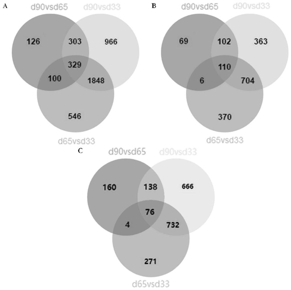
[0058] Example 2 Combined analysis of ChIP-seq and RNA-seq and screening of differentially expressed genes

[0059] 1. ChIP-seq experiment

[0060] (1) Preparation of working reagents

[0061] The working reagent preparation system used in the ChIP-seq experimental process is as follows:

[0062] ①Douncing Buffer (1mL): add 10μl Tris-HCl (pH=7.5), 4μl MgCl2, 2μl CaCl2, 10μl PIC, 974μl ddH2O respectively;

[0063] ②IP Buffer (1 mL): add 10 μl Tris-HCl (pH=8.0), 10 μl Triton X-100, 10 μl Deoxycholate (DOC), 10 μl SDS, 18 μl NaCl, 4 μl EDTA, 10 μl PIC, 928 μl ddH2O;

[0064] ③Hypotonic Lysis Buffer (1 mL): add 0.4 μl EDTA (pH=8.0), 0.1 μl Benzamidine, 1 μl PMSF, 3 μl DTT, 10 μl PIC, 985.5 μl ddH2O respectively;

[0065] ④ChiP Wash Buffer (1mL): add 20μl Tris-HCl (pH=8.0), 10μl SDS, 10μl Triton X-100, 4μl EDTA, 30μl NaCl, 10μl PIC, 916μl ddH2O respectively;

[0066] ⑤ChiP Final Wash Buffer (1mL): add 20μl Tris-HCl (pH=8.0), 10μl SDS, 10μl Triton X-100, 4μl EDTA, 100μl NaCl, 10...

Embodiment 3

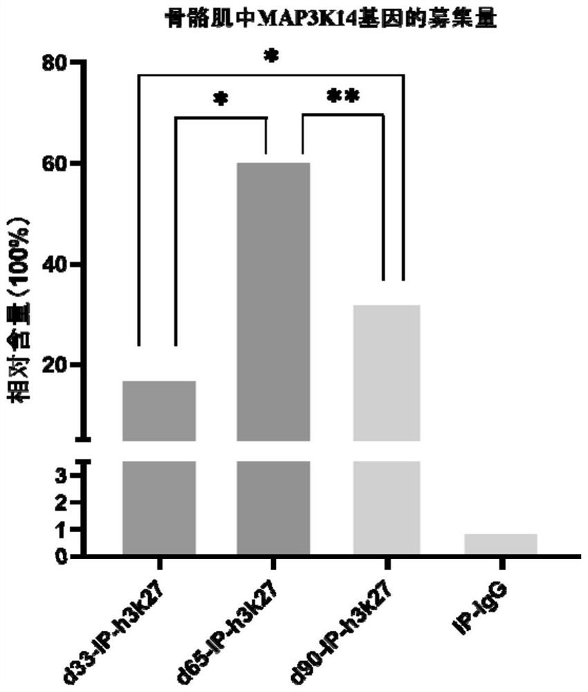
[0157] Example 3 ChIP-qPCR verification of differentially expressed gene MAP3K14

[0158] The quantitative PCR primers for porcine genes were designed using the primer design software Oligo 7.56 as shown in Table 2. Dilute the IP samples incubated with antibodies obtained during chromatin immunoprecipitation to the same concentration, and dilute the Input of 9 samples to 10ng / mL, 5ng / mL, 2.5ng / mL, 1.25ng / mL, 0.625 ng / mL, different concentrations of Input were used to draw a standard curve, and all samples were prepared according to Table 3. Amplification system. Absolute quantitative PCR was performed using illumina's EcoTM Real-time PCR instrument. Each IP sample and different concentrations of Input were used for 3 biological repetitions. The reaction conditions were: 95°C, 5min; 95°C, 10s, 60°C, 15s, 72 ℃, 20s, 40-50 cycles; 95℃, 15s; 60℃, 15s; 95℃, 15s.

[0159] Table 2 ChIP-qPCR primers

[0160]

[0161]Table 3 ChIP-qPCR reaction system

[0162]

[0163] The res...


PUM


Abstract

The invention discloses a method for promoting skeletal muscle development, which comprises overexpressing MAP3K14 gene at embryonic level. Thus, not only was a key target gene MAP3K14 that was modified by H3K27me3 in the development of porcine skeletal muscle discovered, but the enrichment level of this gene was first up-regulated and then down-regulated under the modification of H3K27me3 in the process of porcine skeletal muscle myofiber formation, And it can promote the proliferation function of pig skeletal muscle satellite cells and then promote the development of pig skeletal muscle; it can also promote the development of pig skeletal muscle by overexpressing the MAP3K14 gene, thus providing a new method for improving the lean meat rate of pigs and targets, as well as new research directions; moreover, the application of overexpression of MAP3K14 gene in promoting skeletal muscle development can provide new ideas and targets for lean pig breeding; in addition, in the embryonic stage of pigs By overexpressing the MAP3K14 gene, the effect of promoting skeletal muscle development can be achieved through various methods, and has wide applicability.

Description

technical field [0001] The invention relates to the field of biotechnology, in particular to a method and application for promoting skeletal muscle development. Background technique [0002] Histone methylation is a modification of the N-terminal peptide chain of each subunit of the nucleosome core histone. Among the four subunits that make up the nucleosome, the lysines at the 4th, 9th, 27th, 36th, and 79th positions of the N-terminal peptide chain of the H3 subunit are methylation hotspots, and the methylation types include one, 2. Trimethylation (mono-, di-, tri-methylation). H3K27me3 is the trimethylation of lysine 27 in histone H3 subunit, which mainly plays the role of transcriptional repression and participates in the developmental regulation of skeletal muscle. [0003] The growth and development of porcine skeletal muscle is a rather complex process, mainly including the formation and proliferation of muscle cells, the formation of myotubes and muscle fibers, and ...

Claims


Application Information

Patent Timeline
Patent Type & Authority Patents(China)
IPC IPC(8): C12N15/877C12N15/113C12N5/10C12N15/12A01K67/027
CPCC07K14/47C12N15/8778C12N5/0659C12N15/113A01K67/0275C12N2510/00C12N2310/141A01K2217/05A01K2227/108A01K2267/02
Inventor 吴珍芳顾婷谭宝华周健甘炎民邢萍萍乔佳鑫严冠豪蔡更元李紫聪洪林君杨杰郑恩琴黄思秀徐铮
Owner SOUTH CHINA AGRI UNIV
Learn More