Artificial cardiac muscle tissue fibrosis model, and preparation method, preparation device and application thereof

A technique for tissue fibrosis and myocardium, applied in the field of biomedical engineering, can solve problems such as lack of myocardial infarction drugs in fibrosis models, research and development of human myocardial regeneration, and other problems, and achieve the effect of high-throughput drug testing

Active Publication Date: 2020-09-29
HUBEI UNIV
View PDF3 Cites 3 Cited by
  • Summary
  • Abstract
  • Description
  • Claims
  • Application Information

AI Technical Summary

Problems solved by technology

[0005] Aiming at the problems existing in the prior art, the present invention provides an artificial myocardial tissue fibrosis model, a preparation method and its application, with the purpos

Method used


Image

  • Artificial cardiac muscle tissue fibrosis model, and preparation method, preparation device and application thereof
  • Artificial cardiac muscle tissue fibrosis model, and preparation method, preparation device and application thereof
  • Artificial cardiac muscle tissue fibrosis model, and preparation method, preparation device and application thereof

Examples


Embodiment 1

[0046] Example 1 Preparation of cardiomyocytes from pluripotent stem cells

[0047] The cells used in this example are human induced pluripotent stem cells or human embryonic stem cells, which are induced to differentiate into cardiomyocytes by using small molecules to regulate the WNT signaling pathway in stages, and the specific operations are as follows;

[0048] Human pluripotent stem cells were seeded into 6-well plates coated with Matrigel (Corning) at a seeding density of 3.0×10 6 cells / mL / well, using mTeSR medium (STEMCELL Technologies) in 5% CO 2 1. Cultivate in a constant temperature incubator (Thermo) at 37°C for 3 days until the cells grow to 70%-80% full. To prevent cell death, 10 μM Y27632 (Tocris) was added on the first day of culture.

[0049] Aspirate the old medium and replace it with cell culture medium I to initiate differentiation, in 5% CO 2 , and cultured in a 37°C constant temperature incubator (Thermo) for 2 days. Afterwards, discard the old medium...

Embodiment 2

[0054] Example 2 Construction of engineered myocardial tissue sheet

[0055] Using natural extracellular matrix fibrinogen and Matrigel as the skeleton structure between cardiomyocytes, and then constructing engineered myocardial tissue, the specific operation is as follows:

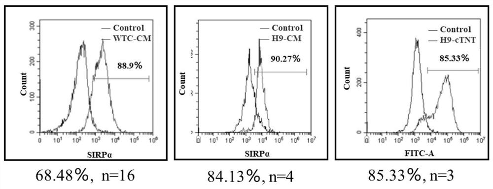
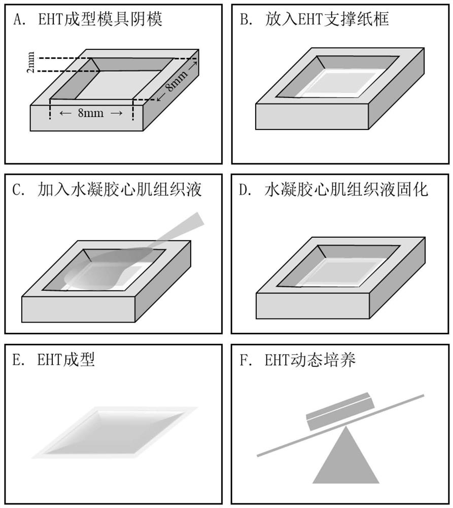
[0056] Use a fused deposition 3D printer (Aurora Corvo) to print the positive mold of the EHT molding mold with polylactic acid (PLA) as the material. Weigh 72 grams of PDMS base solution and add 8 grams of supporting cross-linking agent (SYLGARD TM 184 Silicone Elastomer Kit) was fully stirred and mixed, poured into a glass jar with the obtained male mold, and cured PDMS at 65°C for 4 hours. Separate the male mold, remove the excess cured PDMS, and leave the female mold of the EHT molding mold, such as figure 2 As shown in A. The negative mold was sterilized by high-pressure steam at 121°C for 20 minutes, and the PDMS negative mold was infiltrated with 1‰ F-127 for 30 minutes to increase the hydroph...

Embodiment 3

[0065] Example 3 Freezing injury and self-repair of engineered myocardial tissue slices

[0066] Use a 3D printer (Photon) to make a damage device with light-cured resin, such as Figure 4 A, specifically, a columnar injury device with a pointed end set on the base. Use a 3D printer to make a positive model of the damaged base with light-cured resin.

[0067] Weigh 72 grams of PDMS stock solution, add 8 grams of cross-linking agent, stir and mix well, pour it into a glass cylinder with a male mold of the damaged base, and cure PDMS at 65°C for 4 hours. Separate the male mold of the damaged base, remove the excess cured PDMS, and leave the female mold of the damaged base, such as Figure 4 b. Sterilize the female mold with the damaged bottom at 121°C for 20 minutes by high-pressure steam, wash it with sterile water three times, dry it completely, and set it aside for use.

[0068] Pour the PDMS into the wells of the 24-well plate, cover the bottom, and cure at 65 °C. The o...


PUM


Abstract

The invention belongs to the technical field of biomedical engineering, and particularly relates to an artificial cardiac muscle tissue fibrosis model, and a preparation method, a preparation device and an application thereof. The application provides a first artificial cardiac muscle tissue fibrosis model based on human cardiac muscle cells. On one hand, through a tissue engineering technique, humanized artificial cardiac muscle tissue having human body myocardium structure characteristics is constructed, and after uniform and standardized cryodamage treatment, a fibrosis phenotype which is similar to that after miocardial infarction of a human body and is represented by scarring regions produced due to fiber forming cell activation and extracellular matrix deposition can be generated andmake response to medicines; and on the other hand, due to a mature hiPSC-CM disintegration system and a cardiac muscle tissue construction system, the artificial cardiac muscle tissue fibrosis modelcan be constructed at a low cost in batches, test requirements mainly using 96-hole plate for medicine screening are met, the purpose of high-flux medicine test is achieved, and the dilemma that an existing medicine screening system does not have an artificial cardiac muscle fibrosis model for performing related medicine research and development can be avoided.

Description

technical field [0001] The invention belongs to the technical field of biomedical engineering, and in particular relates to an artificial myocardial tissue fibrosis model, a preparation method, a preparation device and an application thereof. Background technique [0002] Ischemic heart disease is responsible for approximately 9 million deaths per year and has become the leading cause of death worldwide. After heart ischemic infarction, due to the limited regenerative ability of cardiomyocytes in the adult heart, immune infiltration promotes the proliferation, activation and migration of fibroblasts around the infarcted area, and at the same time secretes extracellular matrix to induce pathological remodeling of the infarcted area. This process is called myocardial fibrosis. The intractable scar tissue formed by myocardial fibrosis does not have electrical-contraction properties, which leads to a decline in cardiac function and clinical manifestations of arrhythmia or heart...

Claims


Application Information

Patent Timeline
IPC IPC(8): C12N5/077C12N5/0735C12Q1/02A61K45/00A61P9/00C12M3/00C12M1/24
CPCC12N5/0657G01N33/5061A61K45/00A61P9/00C12M21/08C12M23/06C12M23/12C12M23/48C12M25/14C12M35/00C12N2506/02C12N2501/415C12N2533/56C12N2533/90C12N2501/727C12N2500/38C12N2500/32C12N2503/02G01N2500/10
Inventor 张冬卉杨鹏程龚吉星
Owner HUBEI UNIV
Why Eureka
Try Eureka