Hemostasis scalpel for assisting in cutting treatment through targeted fluorescence imaging

A technology of optical imaging and scalpel, applied in the field of hemostatic scalpel

Pending Publication Date: 2020-10-20
GENGUAN INTELLIGENT TECH HANGZHOU CO LTD
View PDF0 Cites 2 Cited by
  • Summary
  • Abstract
  • Description
  • Claims
  • Application Information

AI Technical Summary

Problems solved by technology

[0005] Today, surgical resection is still one of the effective means of treating most tumors. However, after tumor resection, there is still a risk of tumor regrowth and recurrence because it is difficult to ensure that micro-tumors are removed.

Method used


Image

  • Hemostasis scalpel for assisting in cutting treatment through targeted fluorescence imaging
  • Hemostasis scalpel for assisting in cutting treatment through targeted fluorescence imaging

Examples


Embodiment 1

[0030] Attached below figure 1 A kind of embodiment of the present invention is described in further detail:

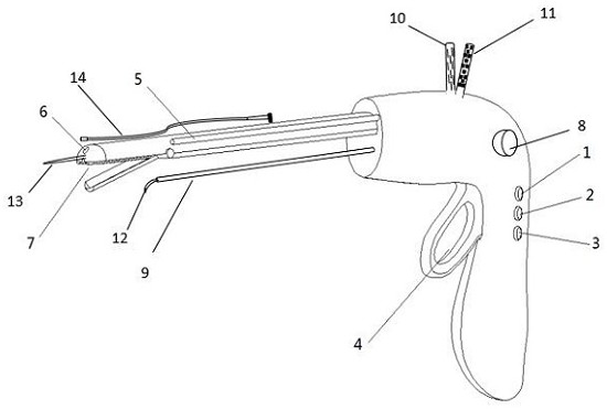
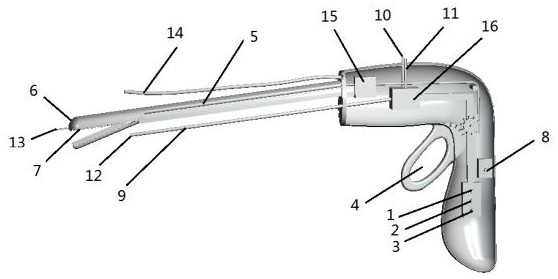
[0031] Such as figure 1 As shown, an embodiment of a hemostatic scalpel for precise targeted cutting and treatment assisted by targeted fluorescence imaging, including a laser switch 1, a cutting gear 2, a hemostatic gear 3, a jaw opening and closing control module 4, and a catheter Channel 5, optical fiber 6, pliers 7, spray gear 8, conduit channel 9, storage tube I10, storage tube II11, atomizing nozzle 12, suction head 13, endoscope 14, ultrasonic energy module 15, transfer pump 16.

[0032] Antibody analogs (which can be proteins, polypeptides, nucleic acids, etc.) modified by upconversion materials with specific recognition of diseased cells that need to be cleaved are formulated in a buffer, and the hose transports the buffer containing the modified antibody analogs to the coupling Catheter channel 5 on the side wall of the operation, and spray on the organ t...

Embodiment 2

[0034] The present invention has hemostatic function, and there are two types of hemostatic methods:

[0035] (1) Use a certain amount of ultrasonic, laser or electric energy to vaporize the water in the bleeding tissue to achieve the purpose of tissue coagulation.

[0036] (2) Apply the hemostatic drug on the bleeding site through the catheter channel 5 .

[0037] In this example, the first method is further described. When the user is in use, when bleeding occurs, press the switch of the hemostatic gear 3 to modulate the energy of the ultrasonic energy module 15 into energy that can coagulate the hemorrhage tissue protein, and use the opening and closing control module 4 of the jaws to control the closing of the jaws 7 to prevent bleeding. tissue for hemostasis.

example 3

[0039] Below in conjunction with accompanying drawing a kind of embodiment of the treatment of the present invention's treatment pathological tissue excision operation is described in further detail:

[0040] Such as figure 1 As shown, an embodiment of spraying the drug-loaded gel on the surgical wound includes a spraying gear 8, a conduit 9, a storage tube I10, a storage tube II11, and an atomizing nozzle 12. When using the spraying device, the spraying gear 8 can be pressed so that the thrombin stored in the storage tube I10 and the collagen solution loaded with tumor suppressor drugs stored in the storage tube II11 quickly pass through the conduit channel 9 at the same time, fully mixed, and pass through The atomizing nozzle 12 is evenly sprayed to the surgical wound at a certain rate to form a gel. The gel also has the effect of stopping bleeding and protecting the wound surface.

[0041] The treatment principles of cancer suppressor drugs are as follows:

[0042] Accor...


PUM


Abstract

The invention relates to a hemostasis scalpel for assisting in cutting treatment through targeted fluorescence imaging. The hemostasis scalpel comprises a laser device, a hemostasis cutting module, acatheter, an endoscope, a suction head and a wound surface treatment module. The catheter sprays up-conversion dye modified targeted antibody to an affected part to be specifically combined with a tumor; the suction head sucks away residual materials; laser is coupled to a knife edge optical fiber head to stimulate dye of the affected part to emit fluorescence; the endoscope displays and observesfocus luminescence; the hemostasis cutting module adjusts output energy to a cutting gear and cuts a luminescence part; the catheter sprays a cleaning solution to an operation area and cleans the operation area; the process is repeated until no visible fluorescence exists; a cancer suppressant is sprayed; and a hemostasis module stops bleeding. The invention aims to realize precise cutting and assist in surgical treatment of tumors.

Description

technical field [0001] The invention belongs to the field of medical devices, in particular to a hemostatic scalpel for accurate cutting and treatment assisted by targeted optical imaging. Background technique [0002] When using traditional surgical methods to treat tumors, because the boundary between diseased tissue and normal tissue cannot be accurately judged, in order to avoid the situation of disease recurrence caused by residual diseased cells, the diseased area is usually judged according to experience, and more patients are used during resection. Part of the normal tissue was resected, and a biopsy was performed to determine whether the resection was complete. However, it takes a long time to judge the effect of surgery by biopsy, and tumor recurrence is likely to occur while waiting for the biopsy results, so part of the normal tissue will be removed during the actual operation. [0003] Optical imaging technology is widely used in biomedicine. There are tumor c...

Claims


Application Information

Patent Timeline
Patent Type & Authority Applications(China)
IPC IPC(8): A61B18/20A61B5/00
CPCA61B5/0071A61B18/20
Inventor 何赛灵余昊明饶杰铭
Owner GENGUAN INTELLIGENT TECH HANGZHOU CO LTD
Why Eureka
Try Eureka