Anti-tumor polypeptide, and preparation method and application thereof

An anti-tumor and anti-tumor drug technology, applied in the biological field, can solve the problems of lack of CLDN18.2 research

Active Publication Date: 2020-10-27
江苏莱森生物科技研究院有限公司
View PDF2 Cites 0 Cited by
  • Summary
  • Abstract
  • Description
  • Claims
  • Application Information

AI Technical Summary

Problems solved by technology

If CLDN18.2 is used as the target, adverse events such as gastrointestinal poisoning can be avoide

Method used


Image

  • Anti-tumor polypeptide, and preparation method and application thereof
  • Anti-tumor polypeptide, and preparation method and application thereof
  • Anti-tumor polypeptide, and preparation method and application thereof

Examples


Embodiment 1

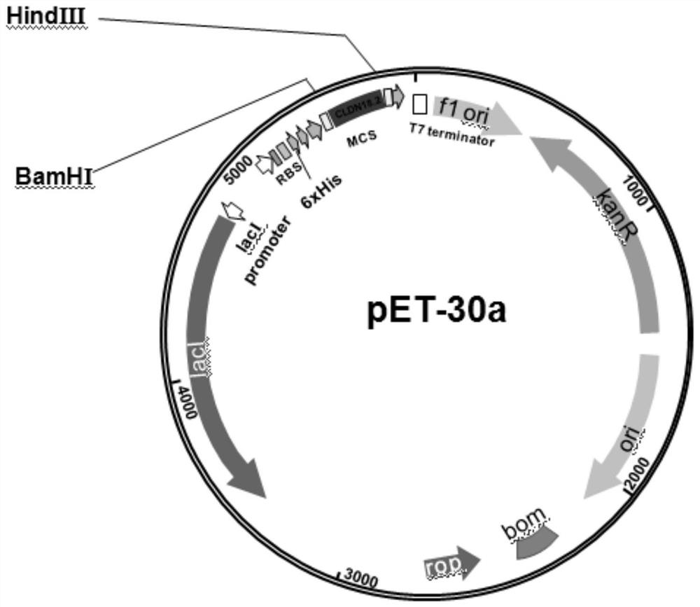
[0018] Example 1: pET-30a / (His) 6 - Construction of CLDN18.2 protein expression vector

[0019] like figure 1 Construct target protein expression vector pET-30a / (His) as indicated 6 -CLDN18.2, search the gene sequence of CLDN18.2 from the Genebank database (1-125 AA, NM_001002026.3), design PCR primers, the upstream primer sequence is: 5'-CG GGATCC ATGGCCGTGACTGCCTGTCAC-3', the downstream primer sequence is: 5'-CC AAGCTT GATGCCTACGATCATCAGGG-3', the underlined part is the restriction site sequence. The PCR product and the vector pET-30a were digested with BamHI and Hind III at 37°C for 3 h, then ligated with T4 DNA ligase at 16°C for 12 h. Transform the ligated product into DH5α competent cells, then spread the transformed product on a kanamycin-resistant (50 μg / ml) LB plate and culture until a single colony grows, pick a single colony, and extract the plasmid for enzyme digestion verification , the recombinant plasmid was sequenced to obtain the recombinant plasmid pET...

Embodiment 2

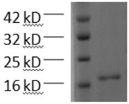
[0020] Example 2: (His) 6 -Expression, purification and validation of CLDN18.2

[0021] The recombinant plasmid pET-30a / (His) prepared in Example 1 6 - CLDN18.2 was transformed into BL21 (DE3) strain, and the recombinant plasmid was screened on a kanamycin-resistant LB plate, and a single colony was picked and cultured in 10 ml LB liquid medium (containing 50 μM kanamycin) until OD 600 After about 0.5, the culture was inoculated in LB liquid medium at a volume ratio of 1: 10, and cultured with vigorous shaking at 37°C until OD 600 About 0.5, add IPTG with a final concentration of 1 mM and induce at 37 °C for 10 h to obtain a bacterial solution.

[0022] Centrifuge the bacterial liquid, remove the supernatant, and separate the bacterial liquid and lysate (50 mM Tris–HCl, 20 mM imidazole, 100 mM NaCl, 10% glycerol, 1% triton, 1 mM protease inhibitor PMSF, 1 mg / ml Lysozyme, pH 8.0) was resuspended in the lysate at a volume ratio of 20:1, placed on ice for 30 min, ultrasonicate...

Embodiment 3

[0024] Example 3. Phage display panning for biologically active peptides that specifically bind to CLDN18.2

[0025] (1) Immobilize the target protein: mix 600 μl of the target protein solution with a concentration of 17 μg / ml (0.1 M NaHCO 3 pH 8.6) into a six-well plate, shake slightly on a shaker, and incubate overnight at 4°C. After washing 6 times with TBST (50 mM Tris-HCl pH 7.5, 150 mM NaCl, 0.1% [v / v] Tween-20), the 3 pH 8.6, 5 mg / ml BSA, 0.02% NaN 3 ) closed for 1 h.

[0026] (2) Screening for specifically binding phages: Wash the six-well plate 10 times with TBST. The amplified phage was diluted with TBST to a copy number of 10 9 ~10 11 In between, the diluted phage was added to the six-well plate to allow it to bind to the target protein, and incubated at room temperature for about 60 min. Wash 10 times with TBST and pat dry after each wash. Add eluent to collect phages that specifically bind to the target protein.

[0027](3) Extraction of monoclonal phage...


PUM


Abstract

The invention belongs to the technical field of biology, and particularly relates to an anti-tumor polypeptide and a preparation method and application thereof. According to the invention, a target gene of an extracellular fragment of CLDN18.2 is introduced into a prokaryotic expression vector, and after induction, a large amount of recombinant protein with a vector tag is expressed. After purification, the relatively pure recombinant protein (His) 6-CLDN18.2 is obtained. Phage in a random dodecapeptide phage library is combined with the recombinant protein, and phage with high affinity is obtained through panning and enrichment. The polypeptide with the highest occurrence frequency is obtained by sequencing and screening after the panned monoclonal phage is amplified and extracted. The anti-tumor polypeptide provided by the invention has remarkable anti-tumor activity and has no acute or chronic toxic effect. The polypeptide provided by the invention has a short sequence and is easy to transport in vivo; a whole polypeptide production process is short in time consumption, low in cost, easy to operate and easy to realize large-scale production, and has wide clinical application value and prospect.

Description

technical field [0001] The invention belongs to the field of biotechnology, and in particular relates to an anti-tumor polypeptide and its preparation method and application. Background technique [0002] The phage display technology is to clone the coding gene fragment of the protein fragment into the appropriate position of the phage coat protein gene, so that the foreign protein fragment and the coat protein are fused and expressed, and the fusion protein is displayed on the surface of the phage with the reassembly of the progeny phage, and the coded this The DNA of the fusion is then located within the virion, allowing a direct link between a large number of random polypeptides and their DNA coding sequences. The displayed protein fragments can maintain a relatively independent spatial structure and bind to target molecules. After a certain period of incubation between the peptide library and the target protein molecules on the solid phase, the unbound free phages are w...

Claims


Application Information

Patent Timeline
IPC IPC(8): C07K7/08C12N15/70C12N15/62C40B30/04A61K38/10A61P35/00
CPCC07K7/08C12N15/70C07K14/705C40B30/04A61P35/00C07K2319/21A61K38/00
Inventor 屠志刚刘晗青
Owner 江苏莱森生物科技研究院有限公司
Why Eureka
Try Eureka