Choroidal OCT image enhancement method and device based on signal reverse compensation

An image enhancement and choroidal technology, applied in the field of OCT, can solve the problems of light signal attenuation, poor signal-to-noise ratio of blood vessels, and difficult segmentation of blood vessel boundaries, etc., to achieve the effect of solving difficult segmentation, enhancing signal-to-noise ratio, and improving visualization and contrast

Inactive Publication Date: 2020-10-30
WENZHOU MEDICAL UNIV
View PDF8 Cites 3 Cited by
  • Summary
  • Abstract
  • Description
  • Claims
  • Application Information

AI Technical Summary

Problems solved by technology

However, the current system does not solve the problem of poor signal-to-noise ratio of deep choroidal vessels caused by backscattering attenuation, which leads to difficulties in vessel boundary segmentation
For this reason, most of the current quantitative choroidal vessel analysis algorithms in OCT images only use traditional image processing methods. However, since the choroid is located below the RPE, the light passes through the RPE and is attenuated by pigment absorption. At the same time, the choroid itself is rich in pigment. The attenuation of the light signal occurs in the depth of the choroid, resulting in blurred boundaries between the choroid and the pigment epithelium and between the choroid and the sclera

Method used


Image

  • Choroidal OCT image enhancement method and device based on signal reverse compensation
  • Choroidal OCT image enhancement method and device based on signal reverse compensation
  • Choroidal OCT image enhancement method and device based on signal reverse compensation

Examples


Embodiment Construction

[0045] The present invention can be better described below in conjunction with the accompanying drawings and some specific embodiments.

[0046] (1) Data acquisition and preprocessing:

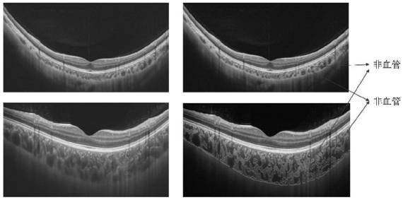
[0047] Obtain fundus images using current commercial instruments or self-built OCT, preprocess the images including appropriate cropping, and retain the OCT choroidal intensity map, as shown in the attached figure 2 shown.

[0048] (2) Reverse attenuation compensation for choroidal signal:

[0049] The signals received by the OCT detector are backscattered and reflected signals. Due to the influence of RPE and choroid’s own pigments on light absorption, the loss of light scattering at a certain wavelength occurs. By extracting the principle and law of choroid scattering light attenuation and constructing a signal compensation and enhancement algorithm, the visualization and contrast of choroid images can be improved.

[0050] The attenuation correction processing algorithm of the OCT signa...


PUM


Abstract

The invention discloses a choroidal OCT image enhancement method and device based on signal reverse compensation. After a choroid image signal acquired by a system is preprocessed, a reverse signal attenuation compensation model is established based on the idea of OCT back scattering signal reverse compensation, the choroid signal is reversely compensated, and the signal-to-noise ratio between a blood vessel and a non-blood vessel tissue is enhanced, so that the defect that the boundary of a choroid blood vessel OCT image is difficult to segment is overcome.

Description

technical field [0001] The invention relates to the field of OCT technology, in particular to a choroidal OCT image enhancement method and device based on signal reverse compensation. Background technique [0002] The choroid is a highly vascularized, pigment-rich tissue located between the retina and sclera. The main function is to provide oxygen and nutrients to the RPE and the outer layer of the retina. The oxygen it transports accounts for about 90% of the oxygen consumption of the retina, which is necessary to maintain the high metabolic activity of the photoreceptor cells in the outer layer of the retina; especially in the fovea without blood vessels area, the choroid is the only way for its material exchange. Many diseases are closely related to abnormal vascular structure of the choroid, such as age-related maculopathy, high myopia maculopathy, diabetic retinopathy and so on. Therefore, it is of great significance to realize the quantitative analysis of choroidal v...

Claims


Application Information

Patent Timeline
Patent Type & Authority Applications(China)
IPC IPC(8): G06T5/00G06T7/12G06T7/136G06T11/00G06N3/04A61B3/12A61B3/10
CPCG06T5/002G06T7/12G06T7/136G06T11/008A61B3/1225A61B3/102G06T2207/10101G06T2207/20081G06T2207/20084G06T2207/20056G06T2207/30101G06T2207/30041G06N3/045
Inventor 周翔天沈梅晓吴昊陈思思金梓
Owner WENZHOU MEDICAL UNIV
Why Eureka
Try Eureka