Vaccine fusion protein

A technology of fusion protein and flagellin, applied in the field of fusion protein to achieve the effect of promoting secretion and correct conformation

Active Publication Date: 2020-11-20
SUZHOU MAXIMUM BIO TECH CO LTD
1 Cites 5 Cited by

AI-Extracted Technical Summary

Problems solved by technology

At present, there is no effective drug for the treatment of the new coronavirus. The development of a targeted...

Method used

[0110] In the present application, the fusion protein may also comprise a CD4+ cell epitope. In the present application, the use of the CD4+ cell epitope can further promote the activation of T cells, thereby promoting the immune response mediated by T cells.
[0117] Fusion proteins described herein may comprise flagellin or fragments thereof. The flagellin described herein may be from Salmonella, Salmonella typhi and/or Filamentous bacteria. In certain instances, the flagellin or fragment thereof may comprise an N-terminal constant region of flagellin and a C-termin...

Abstract

The invention relates to fusion protein. The fusion protein comprises SARS-CoV-2 antigen polypeptide and flagellin or fragments thereof. The invention further provides a preparation method and application of the fusion protein. The fusion protein disclosed by the invention can induce an organism to generate a cellular immune response aiming at an antigen of the SARS-CoV virus.

Application Domain

SsRNA viruses positive-senseViral antigen ingredients +8

Technology Topic

AntigenFlagellin +6

Image

  • Vaccine fusion protein
  • Vaccine fusion protein
  • Vaccine fusion protein

Examples

  • Experimental program(5)
  • Effect test(1)

Example Embodiment

[0175] Example 1 Construction of fusion protein based on flagellin fusion protein
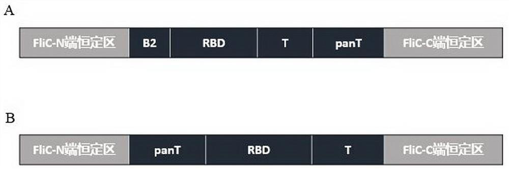
[0176] Construct fusion proteins, from N-terminal to C-terminal: flagellin flagellin (FliC) N-terminal constant region (1-176 amino acids, SEQ ID NO: 6), B cell epitope hot spot region II (B2, SEQ ID NO: 6) NO: 21, SEQ ID NO: 22 and SEQ ID NO: 28 overlap), SARS-CoV-2 receptor binding domain RBD (containing B1 fragment, SEQ ID NO: 5), T cell epitope (SEQ ID NO: 24 overlaps with SEQ ID NO: 25), panT (pan-HLA-DR binding epitope/helper T cell epitope, i.e. CD4 + cellular epitope, SEQ ID NO: 4), and the flagellin FliC C-terminal constant region (amino acids 403-495, SEQ ID NO: 7). Among them, flagellin is an intramolecular adjuvant, which together with the SARS-CoV-2 antigen constitutes a recombinant vaccine, and the obtained fusion protein is called SC2V7 (the amino acid sequence is shown in SEQ ID NO: 9, and the specific structure is shown in figure 2 shown in A).
[0177] The fusion protein was constructed, from N-terminal to C-terminal: flagellin flagellin (FliC) N-terminal constant region (1-176 amino acids, SEQ ID NO: 6), panT (pan-HLA-DR binding epitope/helper T) cell epitope i.e. CD4 + Cell epitope, SEQ ID NO: 4), SARS-CoV-2 receptor binding domain RBD (containing B1 fragment, SEQ ID NO: 5), T cell epitope (SEQ ID NO: 26), and flagellin FliC C-terminal constant region (amino acids 403-495, SEQ ID NO: 7). Among them, flagellin is an intramolecular adjuvant, which together with the SARS-CoV-2 antigen constitutes a recombinant vaccine, and the obtained fusion protein is called SC2V8 (the amino acid sequence is shown in SEQ ID NO: 10, and the specific structure is shown in figure 2 shown in B).
[0178] The nucleotide sequences encoding SC2V7 and SC2V8 are shown in SEQ ID NO: 13 and SEQ ID NO: 14, respectively.
[0179] In addition, the C-terminal constant region of flagellin flagellin (FliC) was replaced by a fragment comprising the amino acid sequence shown in SEQ ID NO: 7 to a fragment comprising the amino acid sequence shown in SEQ ID NO: 8 to construct a fusion without the complete C-terminal Proteins SC2V7-1 (SEQ ID NO: 11) and SC2V8-1 (SEQ ID NO: 12), the nucleotide sequences encoding SC2V7-1 and SC2V8-1 are shown in SEQ ID NO: 15 and SEQ ID NO: 16, respectively. Show.

Example Embodiment

[0180] Example 2 Fusion protein expression verification
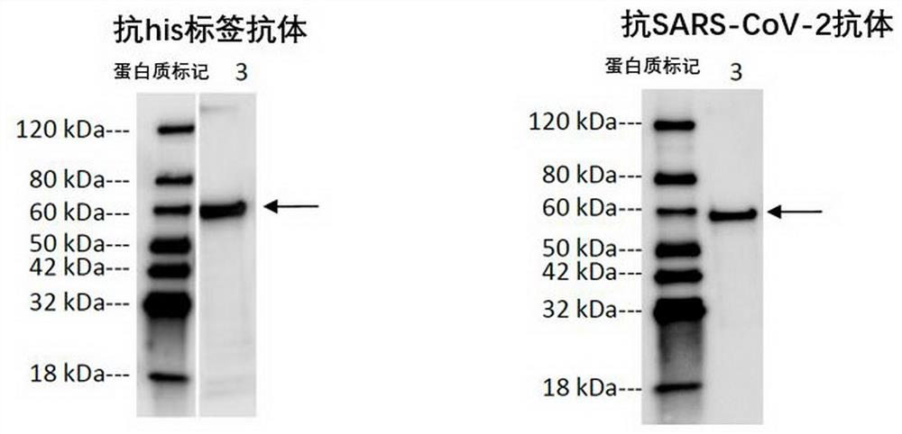
[0181] The nucleic acid sequences encoding SC2V7 and SC2V8 were synthesized by Suzhou Jinweizhi Biotechnology Co., Ltd. and inserted between Nde I and Pst I of the pET21a expression vector (purchased from EMD Biosciences) to obtain the expression vector. The expression vector was transformed into E. coli BL21-Gold (DE3) and grown in LB medium (containing 100 µg/ml ampicillin) overnight at 37°C. Large-scale expression of the transformed E. coli in TB medium (containing 100µg/ml ampicillin), when the DO595 is 2.0-2.5, was induced by adding 1mM IPTG (purchased from Sangon Bioengineering (Shanghai) Co., Ltd.), 37°C , incubate for 4 hours. The cultured bacteria were collected, crushed under high pressure, the bacterial lysate was heated at 42°C overnight, the precipitate was collected by centrifugation, and the precipitate was resuspended in 0.2-0.25M ammonium sulfate solution at 4°C for 2 hours with slow stirring. Centrifuge, the pellet was washed with PBS-Tween buffer (10 mM Na 2 HPO 4 ,1.8 mM KH 2 PO 4 , 137 mM NaCl, 2.7 mM KCl, pH 7.4 and 0.25% Tween20) and resuspended with slow stirring overnight. After centrifugation again, the resulting pellet was resuspended in 3-3.5M urea (PBS, 20% glycerol, pH 9.0) buffer at 4°C overnight. The fusion protein was collected on a size exclusion column (HiLoad 26/60 Superdex 200 pg SECcolumn), the column was equilibrated with 3-3.5M urea (PBS, 20% glycerol, pH 9.0) buffer. Dialyzed overnight at room temperature for reconstitution with 50 mM Tris–HCl, 800 mM NaCl (pH 7.0). The reaggregated fusion protein was further concentrated and purified with 0.5M ammonium sulfate solution. After centrifugation, the pellet was resuspended with PBS-Tween buffer, and then dialyzed against PBS buffer.
[0182] The purified fusion proteins SC2V7 and SC2V8 were obtained. The bacterial solution harvested after IPTG induction and the samples collected by ultrasound were separated by 15% SDS-PAGE and then transferred to PVDF membranes. They were blocked with 1 × TBST containing 5% nonfat milk powder at room temperature for 1 h; -His-tag antibody or anti-SARS-CoV S protein RBD antibody (Sino Biological, cat#40150-T62-COV2), incubate for 1 h at room temperature; wash 3 times with 1× TBST, 5 min each; add HRP-labeled rabbit anti-mouse IgG (1:10000 dilution, purchased from Santa Cruz, USA), incubated at room temperature for 1 h; washed 3 times with 1×TBST for 5 min each, and washed twice with 1×TBS for 5 min each. ECL color development, line negative exposure analysis results. The result is as image 3 and Figure 4 shown, SC2V7 ( image 3 ) and SC2V8 ( Figure 4 ) protein expression was successful and the composition was single.

Example Embodiment

[0183] Example 3 Fusion protein RBD conformation verification
[0184] 1) The correctly folded RBD domain will be able to interact with its receptor ACE2, which is used as a principle to verify the RBD conformation in recombinant vaccines. The recombinant vaccine protein was obtained according to the method of Example 2, and the ACE2 protein (Sino biological, Cat: 10108-H08H) was coated in the ELISA microplate, and after washing, the blocking solution was added to block, and then the recombinant vaccine protein SC2V7 or SC2V8 was added to the microplate. Incubate in the well plate, add anti-RBD antibody (Sino biological, Cat: 40150-T62-COV2) to incubate after washing, add secondary antibody (Jackson ImmunoResearch, Cat: 111-035-003) to incubate after washing, and finally wash and add After the reaction substrate was incubated for an appropriate time, the reaction was terminated, and the data of each well was read to analyze the results. The results showed that both fusion proteins SC2V7 and SC2V8 could interact with ACE2 coated in the microplate ( Figure 5).
[0185] 2) The correctly folded RBD domain induces the animals to produce corresponding antibodies, which can correctly recognize the S1 protein of the new coronavirus (SARS-CoV-2), so as to verify the RBD conformation in the recombinant vaccine.
[0186] Immunized normal C57 mice: 6-8 week old C57 mice were inoculated with the fusion proteins (SC2V7 and SC2V8) prepared in Example 2. In the first week, the immunization dose was 25µg/mouse, and it was halved in the next two weeks. The immune part was injected intramuscularly in the hind leg. After three consecutive weeks of immunization, mice were sacrificed for blood collection and serum separation.
[0187] Immune serum isolation: mice were enucleated or heart blood was collected, and left standing at room temperature for 1 h until the blood coagulated. The coagulated blood was centrifuged at 3000rpm for 10min, and the supernatant was carefully removed to obtain serum. The serum was stored at -20°C after aliquoting.
[0188] Prepare 100mM carbonate coating buffer, dilute the S protein of SARS-CoV-2 virus to 20μg/ml, coat it in ELISA microplate, and incubate at room temperature for 2h. The coating supernatant was discarded, and after washing twice with PBS, the liquid in the plate was carefully removed, blocking solution was added to block, and the cells were incubated at room temperature for 2 h. After washing twice with PBS, immune serum diluted with blocking solution was added and incubated at room temperature for 2 h. After washing 4 times with PBS, appropriate concentration of secondary antibody was added and incubated at room temperature for 1 h. After washing twice with PBS, the reaction substrate was added, incubated at room temperature for 10-20 min, and an equal volume of stop solution was added to terminate the reaction. Read the absorbance data of each sample well and analyze the results.
[0189] The result is as Image 6 As shown, the immune serum after immunizing mice with recombinant vaccine protein SC2V8 (the fusion protein described in this application) can correctly recognize the new coronavirus S protein (SARS-CoV-2).

PUM

no PUM

Description & Claims & Application Information

We can also present the details of the Description, Claims and Application information to help users get a comprehensive understanding of the technical details of the patent, such as background art, summary of invention, brief description of drawings, description of embodiments, and other original content. On the other hand, users can also determine the specific scope of protection of the technology through the list of claims; as well as understand the changes in the life cycle of the technology with the presentation of the patent timeline. Login to view more.

Similar technology patents

Pig feed capable of improving lactation of sows

InactiveCN109418569APromote secretionPromote lactation in sows
Owner:盖树永

Bacillus polymyxa high-yield exopolysaccharide culture medium and application method thereof

ActiveCN113403247Afull of nutritionPromote secretion
Owner:南宁汉和生物科技股份有限公司

Sugar-free cherry fruit jam and preparation method thereof

InactiveCN107788471APromote secretionstimulate appetite
Owner:龙金明

Oral care composition

InactiveUS20180117070A1promote secretionalleviate symptom
Owner:JAPAN MODE

Rhodococcus ruber immunopotentiator and application thereof in vaccine for pigs

PendingCN113975383APromote secretionPromote production
Owner:广东渔跃生物技术有限公司 +1

Classification and recommendation of technical efficacy words

  • Promote secretion

Medicine used for treating tinea pedis and mycosis ungualis

InactiveCN102813703APromote secretionHelp digestion and absorption
Owner:YUECHI SHENLONG SCI & TECH RES ANDDEV CO LTD

Attractant for black pigs

InactiveCN107048005APromote secretionget rid of intestinal gas
Owner:HEFEI LONGYANG AGRI TECH CO LTD

Low-calorie kimchi and production process thereof

InactiveCN104172032Areduce heatPromote secretion
Owner:马国丰

Oral care composition

InactiveUS20180117070A1promote secretionalleviate symptom
Owner:JAPAN MODE

Sugar-free cherry fruit jam and preparation method thereof

InactiveCN107788471APromote secretionstimulate appetite
Owner:龙金明
Why Eureka
Try Eureka