Detection material for anti-leucine-rich glioma inactivated 1 (LGI1) autoantibody in human body fluid and preparation method and application of detection material

A technology of autoantibodies and detection materials, applied in the field of biomedicine, can solve the problems of low sensitivity, cumbersome steps, and large demand for antibodies, and achieve improved sensitivity and specificity, high sensitivity and specificity, and strong operating skills Effect

Inactive Publication Date: 2019-12-24
SHAANXI MYBIOTECH CO LTD
View PDF6 Cites 0 Cited by
  • Summary
  • Abstract
  • Description
  • Claims
  • Application Information

AI Technical Summary

Problems solved by technology

[0005] In view of the problems existing in the prior art, the purpose of the present invention is to provide a detection material for detecting LGI1 autoantibodies, a preparation method and application of the material, and solve the problems of time-consuming detection, cumbersome steps, and antibody demand of the existing detection methods. Large size, high cost, low sensitivity, etc.

Method used


Image

  • Detection material for anti-leucine-rich glioma inactivated 1 (LGI1) autoantibody in human body fluid and preparation method and application of detection material
  • Detection material for anti-leucine-rich glioma inactivated 1 (LGI1) autoantibody in human body fluid and preparation method and application of detection material
  • Detection material for anti-leucine-rich glioma inactivated 1 (LGI1) autoantibody in human body fluid and preparation method and application of detection material

Examples


Embodiment 1

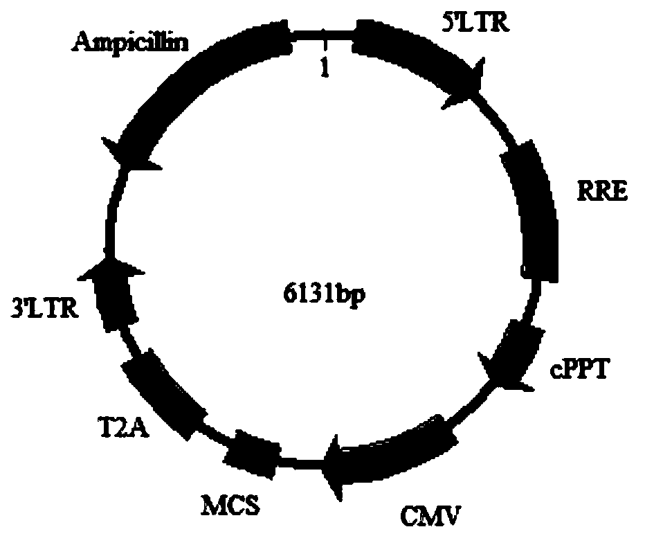
[0038] Step 1, plasmid construction: Obtain the CDS sequence of LGI1 and its signal peptide sequence as the target gene, insert the two target genes with restriction sites into the 17T2A plasmid vector to obtain the recombinant plasmid vector 17T2A-LGI1, and extract it after correct sequencing Plasmid carries out follow-up experiment, and the plasmid vector construction of the present embodiment specifically comprises the following steps:

[0039] Step 1.1: Obtain the CDS sequence of LGI1 and its signal peptide sequence as the target gene by PCR method (artificial synthesis method is also optional), and add SalI / NotI restriction sites at both ends of the two target genes;

[0040]Step 1.2: insert two target genes with restriction sites into the 17T2A plasmid vector, and the insertion site is SalI / NotI to obtain a recombinant vector, which is named 17T2A-LGI1;

[0041] Among them, the 17T2A plasmid vector deletes the copGFP element on the basis of the pCDH-CMV-MCS-EF1-copGFP ve...

Embodiment 2

[0063] This embodiment discloses a method for preparing a sealing material, which specifically includes:

[0064] 1) Cut the carrier film (glass fiber mat in this example) into small square pieces of 0.5cm×0.5cm, put 15μL of a mixture of 1.9M sucrose and 1.9M glucose on each small piece of glass fiber mat, and bake at 100°C for 20min. Store at room temperature for later use;

[0065] 2) Extraction of 17T2A cell (PS) protein: scrape the 17T2A cells obtained in step 2.3 of the above example with a scraper, collect the 17T2A cells in a 15ml centrifuge tube, 1000rpm, 5min, room temperature, discard the supernatant. Add 1mL of 1×PBS (or normal saline) to the precipitate, pipette to mix, transfer the liquid to a 1.5ml EP tube, 700g, 5min, room temperature, discard the supernatant. Add 1 / 2 volume of the cell pellet volume blocking protein extract (1.25% sodium deoxycholate, 0.25% Triton X-100, 0.75% CHAPS, 20mMNaCl, 2×PI), pipette to mix and transfer the liquid to a 2ml EP tube , v...

Embodiment 3

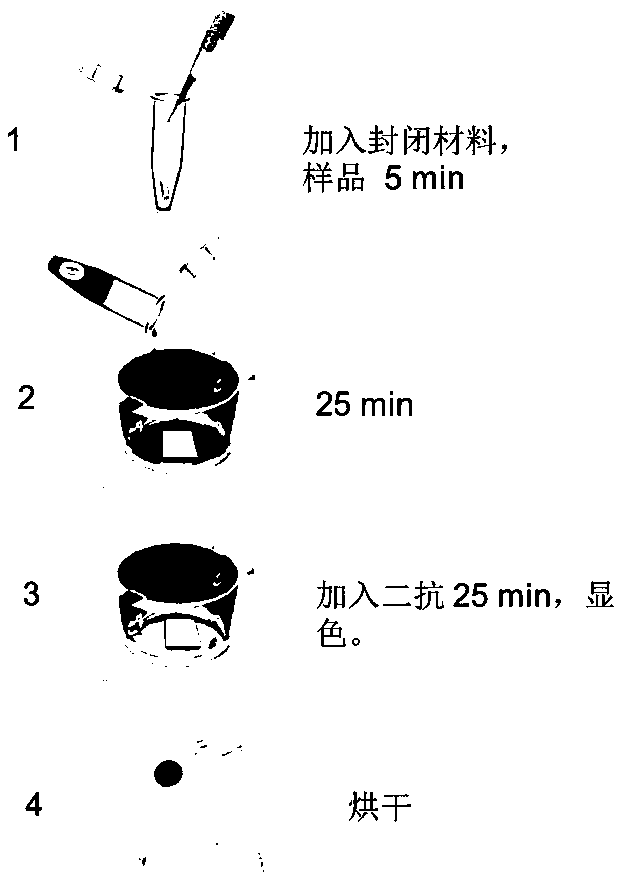
[0069] The detection material for anti-LGI1 autoantibodies in human body fluid prepared in Example 1 is used to detect LGI1 antibodies in samples. Specifically, the detection process for detecting LGI1 antibodies in samples includes:

[0070] 1) Place the LGI1 antigen protein detection material extracted in Step 4 of Example 1 above in a 24-well plate, with the side coated with the membrane protein antigen facing up.

[0071] 2) Serum blocking: the sample to be tested was diluted with working solution (1×PBS, 0.5% Triton X-100, 0.04% EDTA) at a ratio of 1:250, and a piece of blocking material prepared in Example 4 was added to every 250 μL of diluted serum. After standing at room temperature for 2 minutes, vortex for a few seconds, and then stand at room temperature for 5 minutes.

[0072] 3) Serum incubation: Add the blocked serum to a 24-well plate containing membrane protein-cell membrane complex detection material, 250 μL / well, place the 24-well plate on a horizontal shake...


PUM


Abstract

The invention discloses a detection material for an anti-leucine-rich glioma inactivated 1 (LGI1) autoantibody in human body fluid and a preparation method and application of the detection material. The anti-LGI1 receptor detection material is prepared by coating an LGI1 antigen with a carrier membrane, specific LGI1 antibodies in human serum and cerebrospinal fluid are combined with the antigen,color developing is conducted through an alkaline phosphatase substrate-ligand reaction, a sealing material is added to a color developing reaction, and whether an LGI1 antibody is contained in a detection sample or not can be directly judged through observation by naked eyes. The method has the advantages of high sensitivity, easy and convenient operation, rapid detection and the like, and identification and diagnosis of anti-LGI1 receptor encephalitis are facilitated.

Description

technical field [0001] The invention belongs to the technical field of biomedicine, and in particular relates to a detection material, preparation method and application of anti-LGI1 autoantibody in human body fluid. Background technique [0002] Leucine-rich glioma inactivated 1 (LGI1) can regulate the activity of voltage-gated potassium channels and participate in the regulation of neuronal growth and cell survival. Diseases associated with LGI1 include Isaacs' syndrome, epilepsy, familial temporal lobe, autosomal dominant epilepsy with auditory features, and anti-LGI1 autoimmune limbic encephalitis (hereinafter referred to as anti-LGI1 encephalitis). [0003] Meizan Lai et al. tested the blood and cerebrospinal fluid of 57 patients with anti-VGKC encephalitis by immunohistochemistry in 2010, and found that about 41% of the patients detected anti-LGI1 antibodies, so he suggested that anti-VGKC encephalitis Encephalitis was officially renamed anti-LGI1 encephalitis. The s...

Claims


Application Information

Patent Timeline
Patent Type & Authority Applications(China)
IPC IPC(8): C07K14/47C07K1/14C12N15/867G01N33/531G01N33/564
CPCC07K14/4713C12N15/86C12N2740/15043C12N2800/107G01N33/531G01N33/564G01N2333/47G01N2800/28
Inventor 闫亚平李怡婷李科
Owner SHAANXI MYBIOTECH CO LTD
Why Eureka
Try Eureka