
Method for constructing full-retina in-vitro culture model suffering from diabetes retinopathy

A technology of diabetic retina and construction method, which is applied in the field of animal disease models, can solve the problems of ganglion cell apoptosis and short retinal survival time, and achieve good reproducible results

Active Publication Date: 2020-11-24
WEST CHINA HOSPITAL SICHUAN UNIV
View PDF7 Cites 3 Cited by
  • Summary
  • Abstract
  • Description
  • Claims
  • Application Information

AI Technical Summary

Problems solved by technology

Lecleire-Collet A et al reported ganglion cell apoptosis in adult rat retinas cultured in vitro in media containing end-of-glycosylation products for 4 days [Lecleire-Collet A, TessierLH, Massin P, et al. Advanced glycation end products can induce glial reaction and neuronal degeneration in retinal explants[J].Br J Ophthalmol,2005,89(12):1631-1633.], but the survival time of the retina in this study was short, and the test period was only 4 days

Method used


Image

  • Method for constructing full-retina in-vitro culture model suffering from diabetes retinopathy
  • Method for constructing full-retina in-vitro culture model suffering from diabetes retinopathy
  • Method for constructing full-retina in-vitro culture model suffering from diabetes retinopathy

Examples


Embodiment l

[0037] Embodiment 1 Construction of DR model

[0038] 1 Culture medium preparation: the basal medium used for retinal in vitro culture does not contain serum, and is mixed with Neurobasal A, DMEM / F12 culture solution and N2 / B27 supplement at a volume ratio of 50:40:2, and added with a final concentration of 5 mg / L Insulin, 7.5mM D-glucose, 10% (0.1g / ml) BSA and 0.1mM cpt-cAMP were prepared.

[0039] 2 retinal dissection:

[0040] The entire operation must be performed in a sterile environment with all instruments sterilized.

[0041]Ways of killing SD rats at P8: Excessive chloral hydrate or neck dislocation; way of killing embryonic pigs: anesthetized with pentobarbital sodium (intravenous injection about 30 mg / kg) according to body weight, abdominal aorta or femoral artery Euthanasia by exsanguination.

[0042] After the animals were sacrificed, the retinal peeling was carried out according to the following steps: a. Carefully cut off the skin covering the eyeball surface...

Embodiment 2

[0044] Comparison of embodiment 2 with hypertonic control group and normal control group

[0045] 1. Method

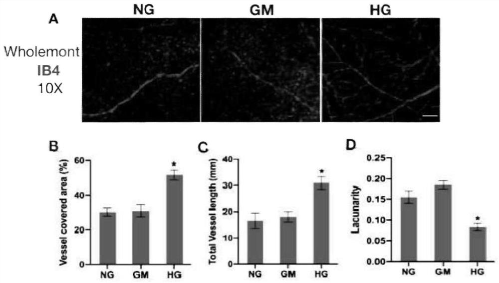
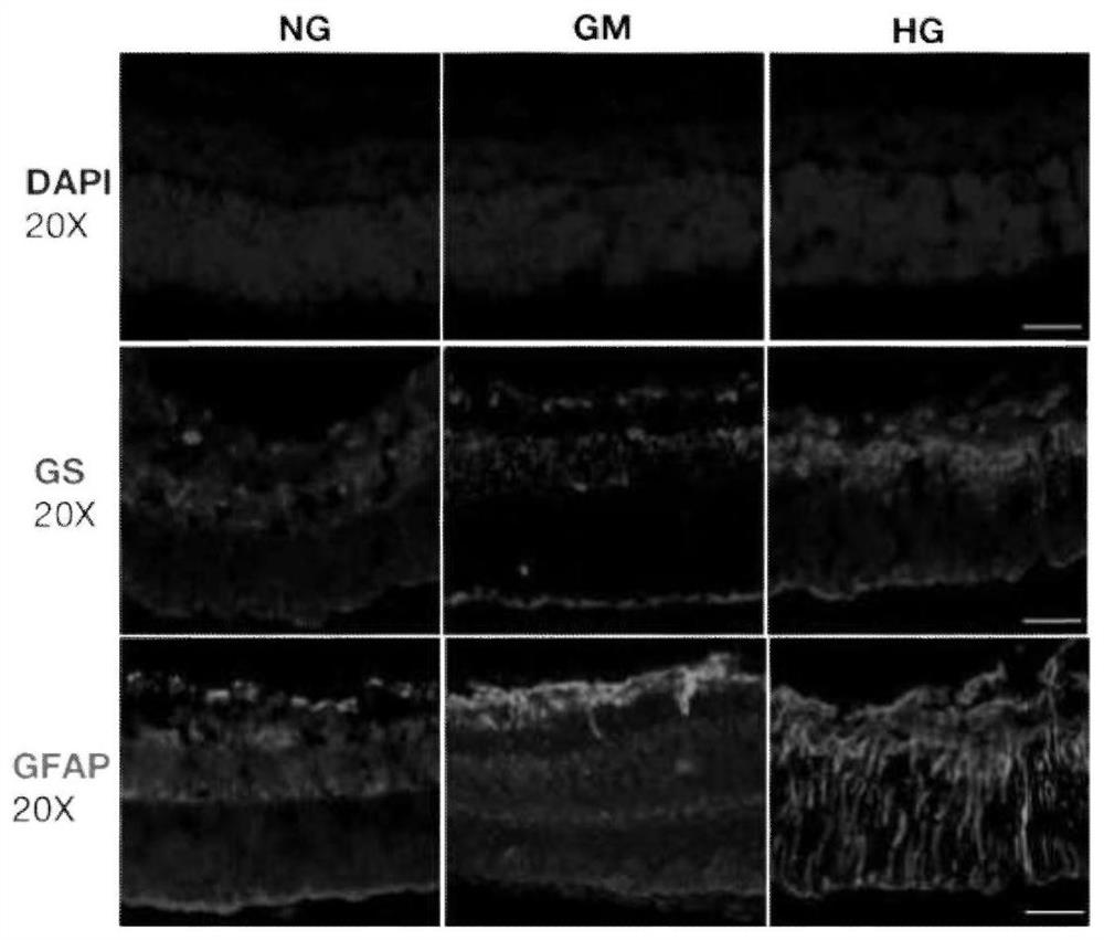
[0046] The retinas of SD rats 8 days after birth (P8) and 90-day embryonic pigs were peeled off according to the method of Example 1, divided into 3 groups, and cultured in three kinds of medium respectively, the temperature of the incubator was set at 37 ° C, CO 2 The content is 5%, cultured for 7 days, and the medium is changed every 2-3 days.

[0047] The medium was prepared as follows:

[0048] High glucose group (HG group): 27.5mmol / L (mM) D-glucose was added to the basal medium (plus the original D-glucose in the basal medium, the final concentration of D-glucose was 35mmol / L);

[0049] Hypertonic control group (GM group): 27.5mmol / L (mM) mannitol was added to the basal medium;

[0050] Normal control group (NG group): basal medium.

[0051] The basal medium is the same as the basal medium in Example 1.

[0052] Immunofluorescent staining was performed on fr...


PUM


Abstract

The invention discloses a method for constructing a full-retina in-vitro culture model suffering from diabetes retinopathy and belongs to the field of animal disease models. The model construction method disclosed by the invention comprises the step of culturing a retina of a rat or fetal pig in a DMEM / F12 culture solution containing D-glucose, Neurobasal A, N2 / B27 supplement, insulin, BSA and cpt-cAMP. According to the method disclosed by the invention, an in-vitro model with typical diabetes retinopathy symptoms can be rapidly obtained, consumed time is short, the method can be repeatable, and the application value is good.

Description

technical field [0001] The invention belongs to the field of animal disease models. Background technique [0002] The global prevalence of diabetes and its complications continues to rise, posing a serious threat to human health. Among them, diabetic retinopathy (DR) is the most common ocular complication of diabetes and the most important cause of blindness in the working-age population worldwide. Since the pathogenesis of DR has not been fully elucidated, its prevention and treatment are still difficult. Microvascular lesions in DR are still the focus of clinical attention. According to the severity of lesions, DR is divided into non-proliferative DR (NPDR) and proliferative DR (PDR (PDR). NPDR is manifested as microvascular tumors, hard exudates, and cotton wool spots. , venous dilatation and hemorrhage, and intraretinal microvascular abnormalities. PDR is characterized by retinal neovascularization; diabetic macular edema, as the main cause of vision loss, can occur in...

Claims


Application Information

Patent Timeline
IPC IPC(8): C12N5/079
CPCC12N5/062C12N2500/34C12N2500/40C12N2501/33Y02A50/30
Inventor 陈大年王钰娇肖丽容
Owner WEST CHINA HOSPITAL SICHUAN UNIV
Learn More