Polypeptide chip and application thereof in virus detection

A polypeptide chip and virus technology, which is applied to a polypeptide chip and its preparation method and its application in the 2019 new coronavirus detection and biomedical fields, can solve the situation that cannot reflect the changes in the severity of the disease course of the patient's immune response to virus infection, and cannot meet the needs of the new type of coronavirus. Issues such as the huge demand for screening and diagnosis of suspected cases of coronavirus infection

Pending Publication Date: 2020-12-04
ACADEMY OF MILITARY MEDICAL SCI
View PDF0 Cites 14 Cited by
  • Summary
  • Abstract
  • Description
  • Claims
  • Application Information

AI Technical Summary

Problems solved by technology

However, some patients with clinical symptoms were also found to be negative for nucleic acid. The reason may be that the RNA is unstable and easy to degrade during sample processing, and the human immune system will clear part of the viral nucleic acid.
In addition, nucleic acid detection cannot reflect the patient's immune response to viral infecti

Method used


Image

  • Polypeptide chip and application thereof in virus detection
  • Polypeptide chip and application thereof in virus detection
  • Polypeptide chip and application thereof in virus detection

Examples


Embodiment 1

[0072] The preparation of embodiment 1 polypeptide chip

[0073] (1) Carry out solid-phase synthesis according to the 968 polypeptides shown in Table 2;

[0074] (2) Select a three-dimensional D-modified glass slide as the substrate of the protein chip, and design the spotting array of the substrate as a 48×42 array;

[0075] (3) Spot all the synthesized polypeptide fragments and positive controls on the surface of the glass slide with a microarray spotter.

Embodiment 2

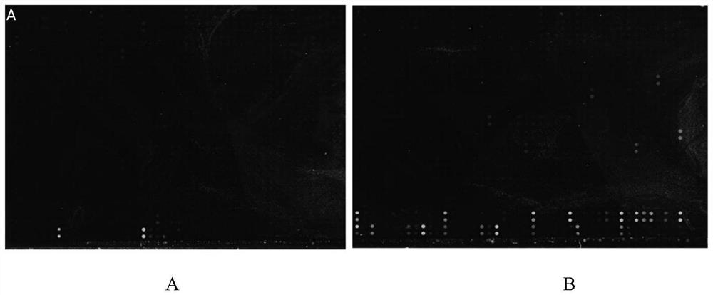
[0076] Example 2 Polypeptide chip detects effectiveness test of SARS-CoV-2 protein antibody

[0077] (1) Sealing: Add a fence to the chip prepared in Example 1, add 400 μl / well 5% milk (0.5g skimmed milk powder + 10ml PBST) to seal at room temperature for 0.5h;

[0078] (2) Sample preparation: the monoclonal antibody of the N protein of anti-SARS-CoV-2, the polyclonal antibody of the N protein of anti-SARS-CoV-2 (both commercially available, original concentration 1mg / ml), add to Mix 400μl 5% milk (1:2000 dilution);

[0079] (3) Adding samples: add the above samples to the chip, 400 μl / well, and incubate at room temperature for 20 minutes;

[0080] (4) Washing: wash 3 times with 0.05% PBST, 5 min each time;

[0081] (5) Add fluorescent dye: Add Goat anti-Rabbit IgG (H+L) Cross-Adsorbed Secondary Antibody, Alexa Fluor 555 (original concentration 2mg / ml, diluted 1:1000 in 400μl 5% milk) to the chip In the array, 400μl / well, incubate at room temperature for 20min in the dark; ...

Embodiment 3

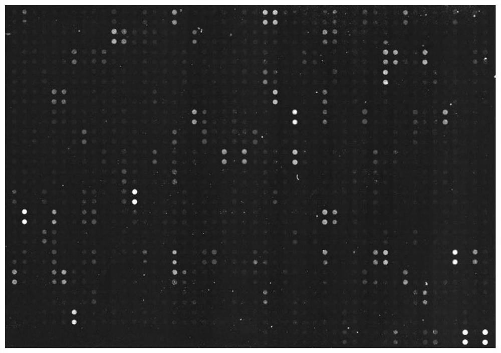
[0085] Example 3 Polypeptide Chip Detects Serum of Patients Infected with SARS-CoV-2 Virus

[0086] (1) Sealing: Add a fence to the chip prepared in Example 1, add 400 μl / well 5% milk (0.5g skimmed milk powder + 10ml PBST) to seal at room temperature for 1 hour;

[0087] (2) Sample preparation: add 4 μl serum to be tested to 400 μl 5% milk and mix well;

[0088] (3) Adding samples: add the detection sample to the chip, 400 μl / well, and incubate at room temperature for 2 hours;

[0089] (4) Washing: wash 3 times with 0.05% PBST, 10 min each time; then wash 3 times with ddH2O, 2 min each time;

[0090] (5) Add fluorescent dye: add goat-anti-hIgG(Fc)-Cy3 (original concentration 1.5mg / ml, 4μl+1200μlmilk) to the chip array, and incubate at room temperature for 1h in the dark;

[0091] (6) Washing in the dark: wash 3 times with 0.05% PBST, 5 min each time;

[0092] (7) Chip detection: dry the chip and detect it with a chip scanner.

[0093] The polypeptide chip of the present in...


PUM


Abstract

The invention provides a polypeptide chip, which comprises a substrate and n polypeptides distributed on the substrate in an array, each polypeptide sequence has 10-20 amino acids, a group consistingof sequences of first to nth polypeptides covers at least 95% of a viral protein sequence, the adjacent polypeptides have an overlap of 3-8 amino acids, and n is 810-1370. According to the method, proteomics and system biology strategies are adopted, all coded protein sequences of COVID-19 are extracted from NCBI data, an SARS-CoV-2 virus proteome polypeptide chip is designed and prepared, and panoramic scanning of all SARS-CoV-2 virus antibodies in blood of a novel pneumonia virus infected patient is achieved.

Description

technical field [0001] The invention belongs to the technical field of biomedicine and relates to a polypeptide chip, in particular to a polypeptide chip and a preparation method thereof and its application in the field of detection of 2019 novel coronavirus (SARS-CoV-2). Background technique [0002] Biochips generally refer to biological information molecules (such as gene fragments, cDNA fragments or polypeptides, proteins, sugars, etc.) fixed on the support medium at high density according to the preset sequence, and realize the biological information based on the principle of specific binding between molecules. Component accurate, fast, high-throughput detection. At present, the common biochips mainly include gene chips, peptide chips, lab-on-a-chip, etc. Among them, peptide chips are getting more and more widely used because of their advantages of being able to directly analyze crude biological samples (serum, urine, body fluid, etc.) and having a wide range of functio...

Claims


Application Information

Patent Timeline
IPC IPC(8): G01N33/68G01N33/58G01N33/577G01N33/569G01N33/552
CPCG01N33/6854G01N33/582G01N33/577G01N33/56983G01N33/552G01N2333/165G01N2469/20
Inventor 于晓波王红叶王聃梁特张晓梅代佳宇
Owner ACADEMY OF MILITARY MEDICAL SCI
Why Eureka
Try Eureka