Water-soluble red fluorescence mitochondria targeting probe and application thereof

A red fluorescence, mitochondrial technology, used in fluorescence/phosphorescence, luminescent materials, material analysis by optical means, etc., can solve the problem of the clarity of the imaging effect, the simplicity of the operation steps needs to be improved, and achieve high signal-to-noise ratio, good biological The effect of compatibility and good signal-to-noise ratio

Active Publication Date: 2020-12-15
HUAZHONG UNIV OF SCI & TECH
View PDF6 Cites 3 Cited by
  • Summary
  • Abstract
  • Description
  • Claims
  • Application Information

AI Technical Summary

Problems solved by technology

The development of fluorescence imaging technology largely depends on the performance of fluorescent materials. AIEgens are widely used in cell imaging due to their high brightness, large Stokes shift and good photostability, as well as excellent biocompatibility. However, the clarity of the imaging effect and the simplicity of the operation steps need to be improved

Method used


Image

  • Water-soluble red fluorescence mitochondria targeting probe and application thereof
  • Water-soluble red fluorescence mitochondria targeting probe and application thereof
  • Water-soluble red fluorescence mitochondria targeting probe and application thereof

Examples


Embodiment 1

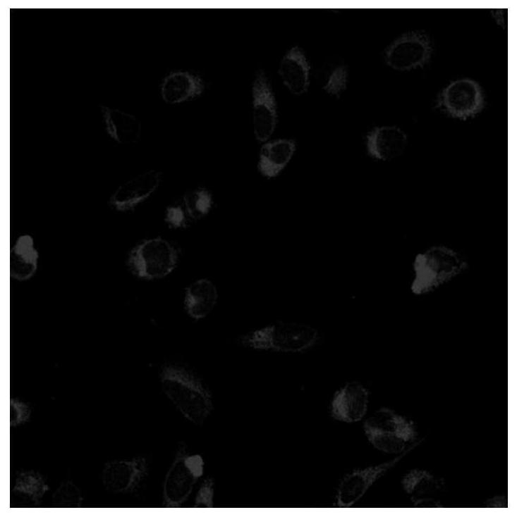
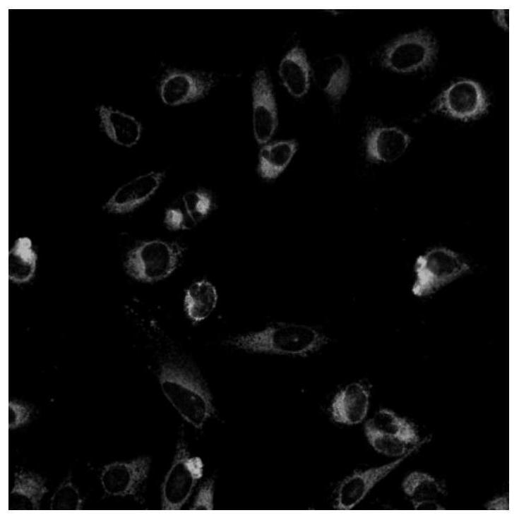
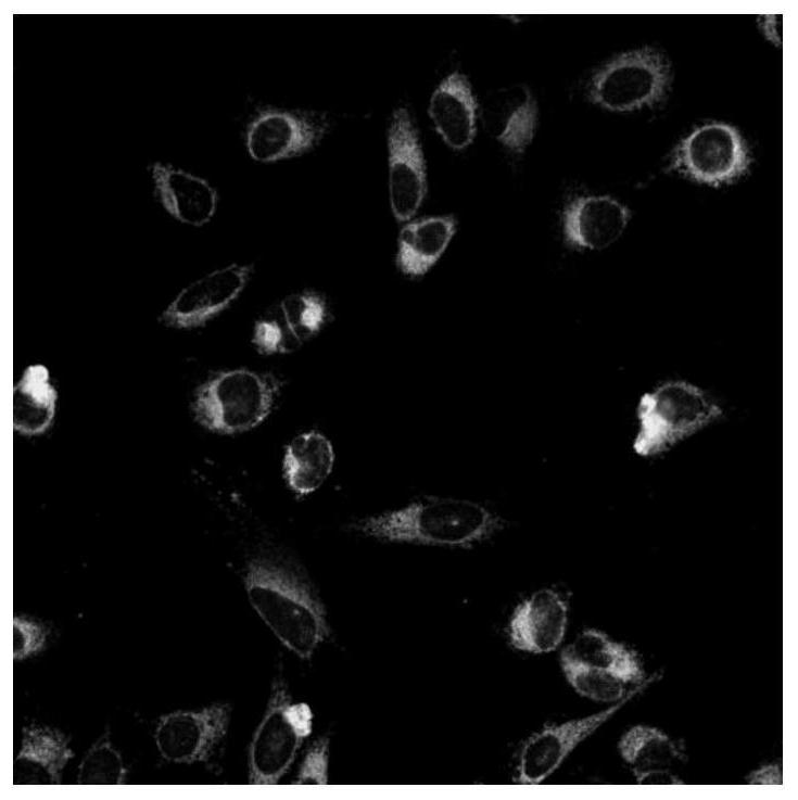
[0061] a structure such as The fluorescent probes shown are abbreviated as TPA-OH. Hela cells were co-stained by TPA-OH and Mito-tracker Green. The concentration of TPA-OH is 2 μM, and the concentration of mitochondrial green fluorescent probe is 100 nM. Fluorescence imaging by confocal microscopy. Finally, a multi-channel fluorescence image is obtained.

[0062] Both the red channel of TPA-OH and the mitochondrial green fluorescent probe (Mito-tracker Green) can see obvious cell outlines, and the images of the two channels are superimposed, as shown in figure 1 The green part and figure 2 The red part in the red channel image of confocal microscope fluorescence imaging overlaps and turns yellow, as shown in image 3 As shown, it is proved that TPA-OH and the mitochondrial green fluorescent probe (Mito-tracker Green) target the same organelle, namely the mitochondria.

Embodiment 2

[0064] a structure such as No. 2 fluorescent probes are indicated. Hela cells were co-stained by fluorescent probe No. 2 and mitochondrial green fluorescent probe (Mito-tracker Green). The concentration of No. 2 fluorescent probe is 2 μM, and the concentration of mitochondrial green fluorescent probe is 100 nM. Fluorescence imaging by confocal microscopy. Finally, a multi-channel fluorescence image is obtained.

[0065] Both the red channel of fluorescent probe No. 2 and the mitochondrial green fluorescent probe (Mito-tracker Green) can see obvious cell outlines, and the images of the two channels are superimposed, as shown in Figure 4 The green part and Figure 5 The red part in the red channel image of confocal microscope fluorescence imaging overlaps and turns yellow, as shown in Figure 6 As shown, it is proved that TPA-OH and the mitochondrial green fluorescent probe (Mito-tracker Green) target the same organelle, namely the mitochondria.

Embodiment 3

[0067] a structure such as No. 3 fluorescent probes are indicated. Hela cells were co-stained by fluorescent probe No. 3 and mitochondrial green fluorescent probe (Mito-tracker Green). The concentration of the No. 3 fluorescent probe is 2 μM, and the concentration of the mitochondrial green fluorescent probe is 100 nM. Fluorescence imaging by confocal microscopy. Finally, a multi-channel fluorescence image is obtained.

[0068] Both the red channel of fluorescent probe No. 3 and the mitochondrial green fluorescent probe (Mito-tracker Green) can see obvious cell outlines, and the images of the two channels are superimposed, as shown in Figure 7 The green part and Figure 8 The red part in the red channel image of confocal microscope fluorescence imaging overlaps and turns yellow, as shown in Figure 9 As shown, it is proved that TPA-OH and the mitochondrial green fluorescent probe (Mito-tracker Green) target the same organelle, namely the mitochondria.

[0069] Result a...


PUM


Abstract

The invention relates to a water-soluble red fluorescence mitochondria targeting probe and application, and belongs to the field of new materials. The invention discloses a water-soluble red fluorescence mitochondria targeting probe, which has a structural general formula shown in the specification, wherein M is an aromatic ring, and N is a hydrophilic group. Compared with other commercial mitochondrial fluorescence probes, the targeting probe provided by the invention has relatively good water solubility. When the fluorescent probe disclosed by the invention is used for dyeing cells, mitochondria can be targeted; the probe has higher biocompatibility and almost has no background fluorescence, and the weak scattered light and autofluorescence of a biological sample at a red fluorescence waveband are beneficial to reducing background signals, so that the probe has a high signal-to-noise ratio. In addition, the red fluorescence has better penetration depth, has more outstanding advantages when being used for mitochondrial imaging of cells, can be used for observing the mitochondrial imaging process of the cells in situ under the rinsing-free condition, and can track the state of themitochondria of the cells for a long time.

Description

technical field [0001] The invention belongs to the field of new materials, more specifically, relates to a water-soluble red fluorescent mitochondria-targeting probe and its application, especially to a new water-soluble fluorescent probe material based on aggregation-induced luminescent properties and its potential in biological imaging Application of targeted staining to mitochondria. Background technique [0002] Cells are an important component of the human body structure. Different types of cells have different functions, and together they maintain the normal operation of life activities. Human cells have a delicate and complex structure, including organelles, cell membrane, cytoplasm, nucleus and cytoplasmic matrix. The organelles include mitochondria, endoplasmic reticulum, lysosomes, lipid droplets, etc. Different organelles have different functions and participate in the entire metabolic process of the cell. The close coordination, division of labor and cooperati...

Claims


Application Information

Patent Timeline
Patent Type & Authority Applications(China)
IPC IPC(8): C07D213/38C07D239/26C07D417/06C09K11/06G01N21/64
CPCC07D213/38C07D239/26C07D417/06C09K11/06G01N21/6428G01N21/6458C09K2211/1044C09K2211/1037C09K2211/1007C09K2211/1014C09K2211/1029
Inventor 李冲田蕊朱明强王亚龙
Owner HUAZHONG UNIV OF SCI & TECH
Why Eureka
Try Eureka