Photomagnetic nanoprobe with core-point shell structure as well as preparation method and application of photomagnetic nanoprobe

A technology of nano-probe and shell structure, applied in nanotechnology, nanotechnology, nano-optics, etc., can solve the problems of incompatibility of relaxation efficiency and biocompatibility, and achieve excellent up-conversion photoluminescence ability, excellent Effect of Magnetic Resonance Relaxation Capacity

Active Publication Date: 2021-02-02
SUN YAT SEN UNIV
View PDF7 Cites 2 Cited by
  • Summary
  • Abstract
  • Description
  • Claims
  • Application Information

AI Technical Summary

Problems solved by technology

[0006] In order to overcome the defect that the relaxation efficiency and biocompatibility described in the prior art cannot be combined, the present invention provid

Method used


Image

  • Photomagnetic nanoprobe with core-point shell structure as well as preparation method and application of photomagnetic nanoprobe
  • Photomagnetic nanoprobe with core-point shell structure as well as preparation method and application of photomagnetic nanoprobe
  • Photomagnetic nanoprobe with core-point shell structure as well as preparation method and application of photomagnetic nanoprobe

Examples


Embodiment 1

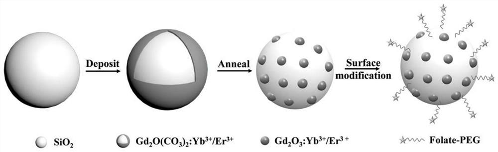
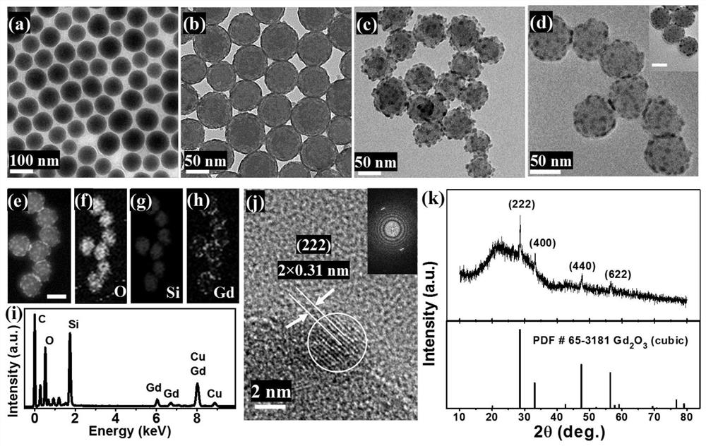
[0054] This embodiment provides a core-point-shell structured optomagnetic nanoprobe, the preparation route is as follows: figure 1 Shown, the preparation method of this optomagnetic nanoprobe is as follows:

[0055] S1. SiO 2 Preparation of nuclei:

[0056] Add 2ml of deionized water to 100ml of absolute ethanol, then add 6ml of 25% (volume fraction) concentrated ammonia water, place it in a constant temperature water bath at 40°C, then add 3ml of tetraethyl orthosilicate, and continue stirring for 24h; Min speed centrifugation for 15min, and then washed 3 times with absolute ethanol and deionized water to obtain SiO 2 nuclei, dissolved in ethanol to give SiO 2 Nuclear ethanol solution and aliquoted into 6 parts for later use.

[0057] S2. Preparation of core-point-shell structured nanoparticles:

[0058] Take 1 part of SiO 2 The nuclear ethanol solution was centrifuged at 10000r / min for 15min to obtain SiO 2 Nuclei, dissolved in 95ml of ionized water containing 3g of ...

Embodiment 2~6

[0063] Embodiments 2 to 6 respectively provide a kind of core-point-shell structured optomagnetic nanoprobe. The difference between the preparation method of the optomagnetic nanoprobe provided in Examples 2 to 6 and Example 1 is that in the metal salt solution in step S2 Yb(NO 3 ) 3 The concentrations are respectively 1mol%, 2mol%, 3mol%, 7mol%, 9mol%;

[0064] The consumption and operation of other raw materials are identical with embodiment 1.

Embodiment 7~11

[0066] Embodiments 7 to 11 respectively provide a kind of core-point-shell structured optomagnetic nanoprobes. The difference between the preparation method of the optomagnetic nanoprobes provided in Examples 7 to 11 and Example 1 is that in the metal salt solution in step S2 Also contains LiNO 3 , LiNO 3 The concentrations are respectively 1mol%, 2mol%, 4mol%, 6mol%, 8mol%;

[0067] The consumption and operation of other raw materials are identical with embodiment 1. The core-point-shell structure nanoparticles prepared in Examples 7-11 are named as SiO 2 @Gd 2 o 3 :Yb 3+ / Er 3+ / Li + nanoparticles.


PUM

PropertyMeasurementUnit
Particle sizeaaaaaaaaaa
The average particle sizeaaaaaaaaaa
Particle sizeaaaaaaaaaa

Abstract

The invention discloses a photomagnetic nanoprobe with a core-point shell structure as well as a preparation method and application of the photomagnetic nanoprobe. The photomagnetic nanoprobe comprises a SiO2 core, Gd2O3 particles which are used as point shells, are distributed on the surface of the SiO2 core and are doped with metal ions, and target molecules which are hung on the surface of theSiO2 core, wherein the metal ions are Yb < 3 + > and Er < 3 + >, or Yb < 3 + >, Er < 3 + > and Li < + >. The photomagnetic nanoprobe with the core-point shell structure provided by the invention has high magnetic resonance relaxation enhancement performance, biocompatibility and structural stability, and can be applied to nuclear magnetic resonance enhanced imaging as a contrast agent; and meanwhile, the photomagnetic nanoprobe has excellent up-conversion photoluminescence capability, and is converted into red light and green light under near-infrared excitation, so that the photomagnetic nanoprobe has high-sensitivity multicolor optical imaging of tumor cells.

Description

technical field [0001] The invention relates to the technical field of nano-biological materials, and more specifically, to a core-dot-shell structured optomagnetic nanoprobe and its preparation method and application. Background technique [0002] Magnetic resonance imaging (MRI) is based on the principle of nuclear magnetic resonance, and a linear gradient field is added to encode the magnetic nucleus in space, so as to obtain the image between the position of the observed nucleus and the corresponding magnetic resonance signal. An important diagnostic tool that is widely used. Magnetic resonance imaging contrast agent is an important part of this technology, which can change some parameters of the imaging nucleus, thereby improving the imaging contrast. [0003] In 1987, Gd-DTPA was officially approved by the U.S. Food and Drug Administration as an MRI contrast agent. A large number of pharmacological and clinical application studies have proved that Gd-DTPA is a safe, c...

Claims


Application Information

Patent Timeline
IPC IPC(8): C09K11/02C09K11/78B82Y20/00B82Y25/00B82Y30/00B82Y40/00A61K49/08A61K49/12A61K49/18G01N21/64
CPCC09K11/02C09K11/7769B82Y20/00B82Y25/00B82Y30/00B82Y40/00G01N21/6428G01N21/6486A61K49/08A61K49/126A61K49/183A61K49/186
Inventor 殷金昌邵元智曲鸿斌汪项张吴忌郑宏挺林晋纬吴中华
Owner SUN YAT SEN UNIV
Why Eureka
Try Eureka