Method for constructing retinitis pigmentosa disease model, application and breeding method
A technology for retinitis pigmentosa and degenerative diseases, applied in the field of building retinitis pigmentosa disease models, can solve the problems of few types of RP disease models, unclear specific molecular mechanisms, and obstacles to clinical diagnosis and treatment.
- Summary
- Abstract
- Description
- Claims
- Application Information
AI Technical Summary
Problems solved by technology
Method used
Image
Examples
Embodiment 1
[0067] 1. Construction of retinitis pigmentosa disease model
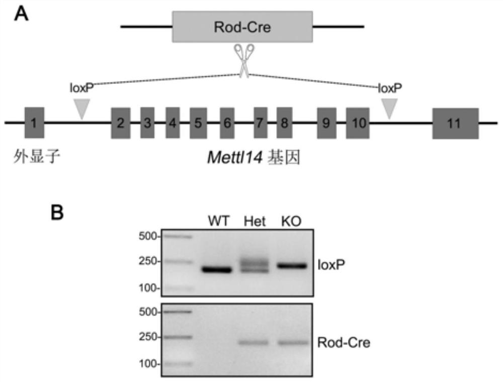
[0068] In this example, mice are used as the target animal, and the method for constructing the retinitis pigmentosa disease model provided by the example of the present invention is described. The route of knocking out the Mettl14 gene is as follows: figure 1 As shown in A, the modification method is: deletion of exons 2-10 of the mouse Mettl14 gene; modification is achieved by combining CRISPR / Cas9 technology and Cre-loxP gene knockout technology. The specific operation is as follows:
[0069] 1) Mettl14 gene conditional knockout mice (Mettl14 em1Flv , the upstream of exon 2 and downstream of exon 10 of the mouse Mettl14 gene are inserted with loxP sites in the same direction) donated by Professor Richard Flavell of Yale University;
[0070] 2) mating and breeding the Mettl14 gene conditional knockout heterozygous mice obtained in step 1) to obtain Mettl14 gene conditional knockout homozygous mice;
[0071] 3)...
Embodiment 2
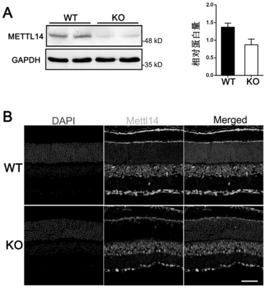
[0095] 1 Western blot analysis of the gene knockout efficiency in the retina of Rod-Cre knockout mice.
[0096] method:
[0097] 1) Obtain the retinas of control and knockout mice respectively, and add 200 μl protein lysate RIPA after thorough grinding.
[0098] 2) After sonicating the cells, they were lysed on ice for 20 min.
[0099] 3) After centrifugation at 16,000 g for 10 minutes at 4°C, transfer the supernatant to another clean centrifuge tube, add 50 μl of protein sample solution, mix well and heat at 95°C for 5 minutes.
[0100] 4) After the samples are cooled, take 20 μl respectively and perform polyacrylamide gel electrophoresis (SDS-PAGE) at 160V to separate proteins.
[0101] 5) After the SDS-PAGE is finished, cut the nitrocellulose membrane of appropriate size according to the needs, lay filter paper, glue, nitrocellulose membrane and filter paper in order, and drive away the air bubbles, put the transfer membrane tank into the ice water bath, and use A consta...
Embodiment 3
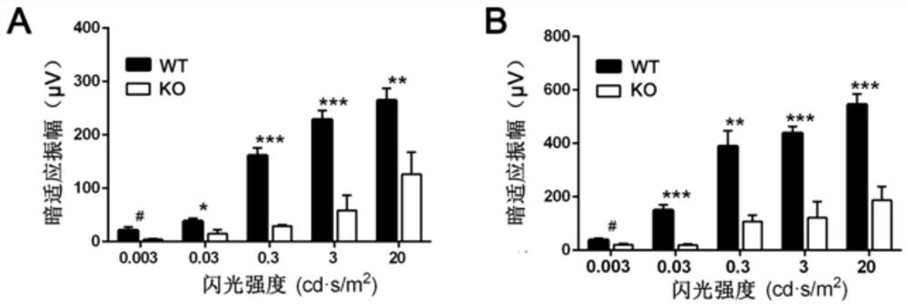
[0111] ERG visual acuity detection of 5-month-old Mettl14 knockout mice:
[0112] 1) Dark-adapted animals should be dark-adapted all night, and the environment should be absolutely dark;
[0113] 2) Anesthesia the next day: weighing, intraperitoneal injection; deep anesthesia is appropriate;
[0114] 3) Animal immobilization and mydriasis: After the anesthesia is completed, fix the mouse with adhesive tape in front of the animal test platform under dark red light illumination: it is necessary to ensure that the mouse is "lying", that is, relative to the stimulation port of the flash stimulator, The eyes are at the same height, fully exposed, and mydriatics are added.
[0115] 4) Electrode installation: After preheating the electroretinograph (Espion Visual Electrophysiology System, Diagnosys LLC, Lit-tleton, MA, USA), apply conductive paste to the electrode, clamp the tail of the mouse, and insert it into the "ground" interface of the amplifier; The double-headed needle elec...
PUM
 Abstract
Description
Claims
Application Information
 - R&D
- Intellectual Property
- Life Sciences
- Materials
- Tech Scout
- Unparalleled Data Quality
- Higher Quality Content
- 60% Fewer Hallucinations
Browse by: Latest US Patents, China's latest patents, Technical Efficacy Thesaurus, Application Domain, Technology Topic, Popular Technical Reports.
© 2025 PatSnap. All rights reserved.Legal|Privacy policy|Modern Slavery Act Transparency Statement|Sitemap|About US| Contact US: help@patsnap.com


