Method for establishing simulated valvular stromal cell calcification model

A technique for establishing a method and valve, applied in the field of biomedicine, which can solve the problems that the calcification pathways are not very related, and it is difficult to study the relationship in detail.

Active Publication Date: 2021-04-30
WEST CHINA HOSPITAL SICHUAN UNIV
View PDF4 Cites 4 Cited by
  • Summary
  • Abstract
  • Description
  • Claims
  • Application Information

AI Technical Summary

Problems solved by technology

Although several calcification-inducing media currently reported can induce calcification in VICs, they simplifies the influence of each factor on CAVD, and it is difficult to study the relationship between each factor in detail, so that the research conclusions based on this may be inconsistent with The true calcification pathway is less relevant

Method used


Image

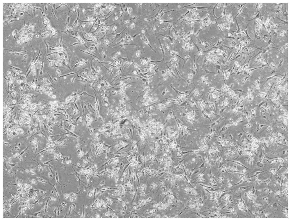
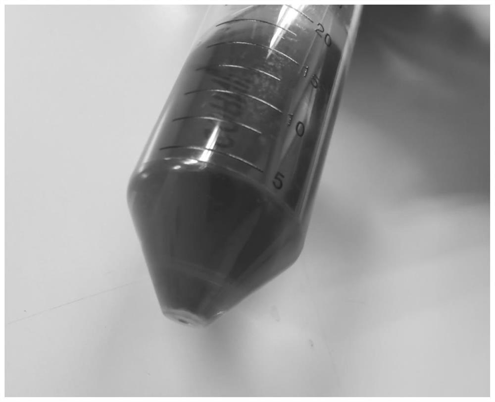
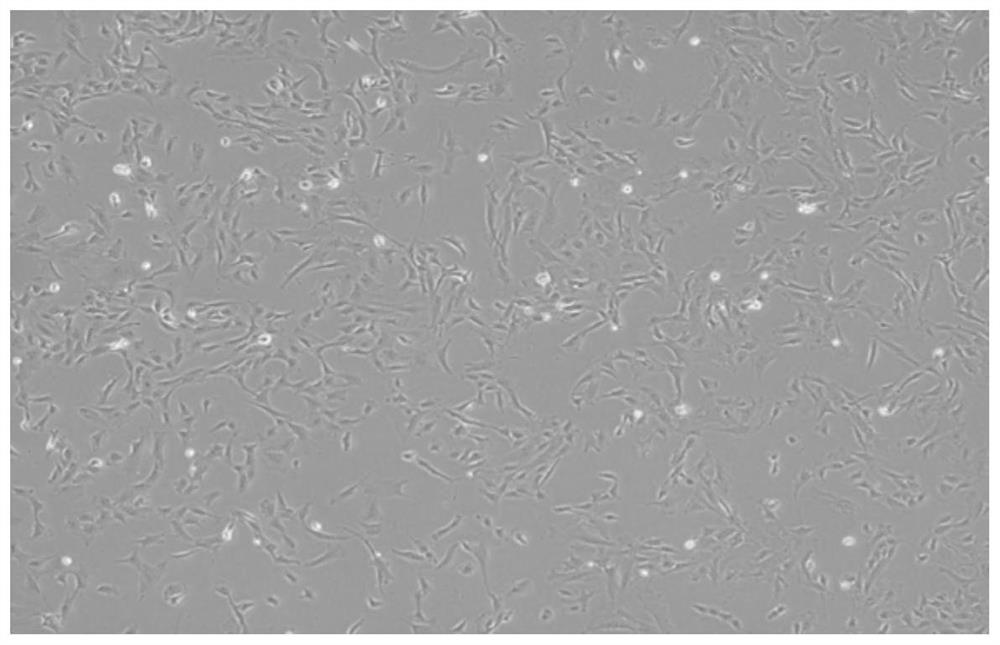
  • Method for establishing simulated valvular stromal cell calcification model
  • Method for establishing simulated valvular stromal cell calcification model
  • Method for establishing simulated valvular stromal cell calcification model

Examples


Embodiment 1

[0056]Example 1 and Example 2 The reagents and arrangements are as follows:

[0057]1: 50 ml of phosphate buffered saline, PBS, no calcium magnesium, Gibco, C10010500BT) + 1% penicillin-streptomycin solution (100X, Biyun Tian, ​​C0222);

[0058]2: 15 ml of 2 mg / ml of type II collagen enzyme digestion liquid: 15 mL PBS + 30 mg Type II, Worthington, LS004176);

[0059]Reagent 3: 100 ml complete medium: 84 ml DMEM high sugar medium (Dulbecco's Modified Eaglemedium, Gibco, C11995500BT) + 15ml fetal bovine serum, FBS, Gibco, 10099141C) + 1ml 1% penicillin-streptomycin solution ;

[0060]Reagent 4: 100 ml cell digestion (GIBCO, 0.25% Trypsin-EDTA, 2186972);

[0061]Reagent 5: 100 ml of condition induced medium: 84 ml reagent 3 + 15ml fetal bovine serum + 1% penicillin-streptomycin solution;

[0062]The reagent 6: 20 ml 1.5 mg / ml of type II collagen enzyme digestion: 20 mL PBS + 30 mg II collagenase (Worthington, LS004176).

[0063]Example 1

[0064]The present invention provides a method for establishing a c...

Embodiment 2

[0093]The present invention provides a method of establishing a calcification model of a simulated valve interstitial cells for human VICS simulation calcification model, including the following steps:

[0094]S1 Prepare for the original calcified VICS

[0095]S1.1 follows my country's relevant laws and regulations to obtain a calcified valve tissue that is no longer needed by patients after CAVD patients, maintains aseptic, and placed in a pre-cooling reagent 1 to transport to the laboratory;

[0096]S1.2 rinse three times in the pre-cooled reagent 1 in S1.1 to remove blood cells, and then place each valve leaflet into a petri dish containing 3 ml reagent 2 (35 mm) In the case of 37 ° C for 30 min;

[0097]S1.3 Wipe the valve lobes in S1.2 in a sterile cotton swab (daily cotton swab high-temperature high-pressure sterilization), to ensure that each part is wiped to remove surface endocytes, will wipe The latter valve was transferred into 13 ml of reagent 2, and the sprinkled digestion overnigh...


PUM


Abstract

The invention discloses a method for establishing a simulated valvular stromal cell calcification model, which belongs to the technical field of biomedicine and comprises the following steps: S1, separating primary calcified VICs; S2, preparing a conditional induction culture medium; S3, performing isolated culture on normal VICs; and S4, performing calcification induction. According to the invention, primary-generation VICs of a human or a rat are induced by a collected primary-generation cell-derived conditioned medium of the VICs of a calcified patient, so that the primary-generation VICs of the human or the rat are converted into osteoblast-like cells from the VICs. Therefore, a set of model capable of reflecting the pathological process of a clinical CAVD patient is established, more favorable conditions are provided for researchers, and the model is good in stability, easy to store, simple to prepare, capable of simulating a clinical pathological model to a greater extent and higher in simulation performance.

Description

Technical field[0001]The present invention relates to the field of biomedical technology, and more particularly to the establishment of a calcified model of simulated valve interstitial cells.Background technique[0002]Calcific aortic valve disease (CAVD) is a pathological process involving lipid deposition, chronic inflammation regulation and progressive calcification, characterized in that the aortic valve leaflet is thickened, the aortic valve ring narrow, left ventricular The increase in mechanical stress causes severe complications. CAVD is the most common cardiac valve disease in the elderly. In 5 years, patients with heart failure, the risk of replacement or death is very high. CAVD is diverse, the pathogenesis is complex, the molecular and cell processes have not been clearly characterized.[0003]However, the primary cellular component valve interstitial cellular components of the aortic cells (VICS), which differs from osteogenous cells (ie, calcification), which is considere...

Claims


Application Information

Patent Timeline
Patent Type & Authority Applications(China)
IPC IPC(8): C12N5/077C12N5/071
CPCC12N5/0654C12N5/0602C12N2509/00
Inventor 李君丽陈茂廖延标黄方洋瞿天一邵若宸
Owner WEST CHINA HOSPITAL SICHUAN UNIV
Why Eureka
Try Eureka