UGT enzyme activity detection method and application thereof

A detection method and technology for enzyme activity, applied in the field of medicine, can solve the problems of low detection throughput, cumbersome and complicated operation, and inability to screen and evaluate multiple UGT subtype enzymes on a large scale, achieving good repeatability, convenient operation, and low cost. The effect of the cost of experiments

Pending Publication Date: 2021-04-30
SHANGHAI UNIV OF T C M
View PDF0 Cites 2 Cited by
  • Summary
  • Abstract
  • Description
  • Claims
  • Application Information

AI Technical Summary

Problems solved by technology

For the activity detection of other UGT subtype enzymes, we can only rely on the traditional non-fluorescent substrate 4-methylumbelliferone (4-MU), which has low detection throughput, and the sample pretreatment and detection process are cumbersome and complicated, and cannot be synchronized. Carry out large-scale screening and evaluation of the inhibitory ability of target substances on various UGT subtype enzymes

Method used


Image

  • UGT enzyme activity detection method and application thereof
  • UGT enzyme activity detection method and application thereof
  • UGT enzyme activity detection method and application thereof

Examples


Embodiment 1

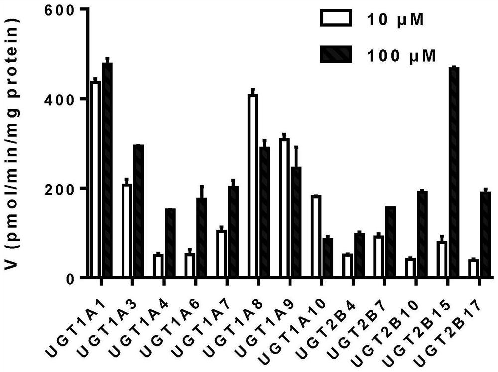
[0037] Example 1. Broad-spectrum research on the metabolism of Ophiopogon japonicus high isoflavone A by human UGT enzyme

[0038]Phenotype study of UGT enzymes involved in glucose-binding metabolism of broad-spectrum substrate Ophiopogon japonicus high isoflavone A. Using 13 commercialized recombinant human UGT single enzymes (UGT1A1, UGT1A3, UGT1A4, UGT1A6, UGT1A7, UGT1A8, UGT1A9, UGT1A10, UGT2B4, UGT2B7, UGT2B10, UGT2B15, UGT2B17) as the target UGT enzyme source, the substrate MOA Metabolic characterization. details as follows:

[0039] (1) Mix Tris-HCl buffer (50mM, pH=7.4) and magnesium chloride (5mM), and use one of the 13 UGT single enzymes as the target UGT enzyme (0.05mg / ml recombinant human UGT single enzyme), Ophiopogon japonicus high isoflavone A (concentration uniformly 10 μM and 100 μM) was mixed in the EP tube, and pre-incubated at 20-60°C for 3-5 minutes, and the pH value of the incubation system was 5-10. Concrete reaction condition is as follows:

[0040]...

Embodiment 2

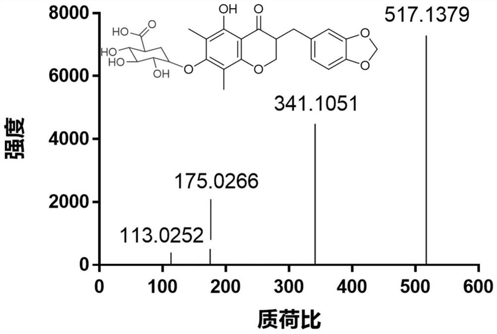
[0045] Example 2. Preparation and identification of Ophiopogon japonicus high isoflavone A metabolites by UGT enzymes

[0046] After using high-resolution mass spectrometry to analyze and identify the metabolites of Ophiopogon japonicus high isoflavone A catalyzed by UGT (such as figure 2 ), a large number of preparations and structural characterizations of its product MOAG were carried out by using biological preparation methods and NMR techniques, respectively. (As shown in Figure 6). Specific steps are as follows:

[0047] (1) Ophiopogon japonicus high isoflavone A (8.52mg), Tris-HCl buffer solution (50mM, pH 7.4), MgCl 2 (5mM), polyoxyethylene hexadecyl ether (0.1mg / mg protein) and 13 recombinantly expressed UGT enzymes (1.85μL for each enzyme, final protein concentration of 0.8mg / ml) were mixed. After 5 min pre-incubation at 37°C, UDPGA (2 mM) was added to start the reaction.

[0048] (2) After incubation at 37°C for 6 hours, the reaction was terminated by adding ice...

Embodiment 3

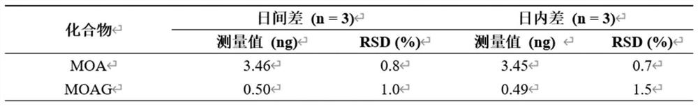
[0052] Embodiment 3. Stability investigation

[0053] (1) Mix Tris-HCl buffer (50mM, pH=7.4) with MgCl under physiological conditions 2 (5mM), with 13 kinds of UGT enzymes expressed recombinantly (0.31 μL for each enzyme, the final protein concentration is 0.1mg / ml) as the enzyme source, and the substrate Ophiopogon japonicus high isoflavone A solution (concentration selection 1 / 10~1K m ) were mixed in a centrifuge tube, the reaction temperature was 37°C, and the pH of the incubation system was 7.4.

[0054] (2) Pre-incubation: incubate the mixture in (1) at 37° C. for 3-5 minutes to ensure sufficient contact between the enzyme and the substrate Ophiopogon japonicus high isoflavone A.

[0055] (3) Add UDPGA and the reaction time is 30 minutes.

[0056] (4) Samples were taken and analyzed after being placed at 4°C for 0 hour, 24 hours and 48 hours to investigate its stability under the storage condition of 4°C refrigerator. The result is as image 3 shown.

[0057] The res...


PUM


Abstract

The invention provides a UGT enzyme activity detection method not used for disease diagnosis and treatment and application of the UGT enzyme activity detection method in screening and evaluating a UGT enzyme regulating agent. According to the detection method, radix ophiopogonis isoflavone A is used as a substrate, and the conversion rate of the radix ophiopogonis isoflavone A or the generation rate of a 7-O-glucuronic acid product of the radix ophiopogonis isoflavone A is quantitatively detected through a liquid-phase ultraviolet method or a liquid-phase mass spectrometry method so as to detect the activity of UGT enzyme. The method disclosed by the invention has the characteristics of high sensitivity, strong stability, good repeatability, simplicity, easiness in operation and the like; the method is used for screening and evaluating various reported UGT enzyme regulators, has extremely high accuracy and has a very good application prospect.

Description

technical field [0001] The invention belongs to the technical field of medicine, and in particular relates to a detection method of UGT enzyme activity not used for disease diagnosis and treatment, and the application of the detection method in the screening and evaluation of UGT enzyme regulators. Background technique [0002] The uridine diphosphate glucuronosyltransferase (UGT) superfamily is an important phase II metabolic enzyme in the body, which increases the hydrophilicity of the substrate by catalyzing the combination of the compound with the cofactor uridine diphosphate glucuronide (UDPGA) Sex, so that the compound can be more effectively excreted in urine or bile, which is an important detoxification process of the body. Mammalian UGTs can be divided into four families: UGT1, UGT2, UGT3 and UGT8. Among them, those involved in drug conjugation and metabolism are mostly members of UGT1A and UGT2B subfamilies. [0003] As the most important phase II metabolic enzym...

Claims


Application Information

Patent Timeline
Patent Type & Authority Applications(China)
IPC IPC(8): C12Q1/48G01N30/02G01N30/74G01N30/72
CPCC12Q1/48G01N30/02G01N30/72G01N30/74G01N2030/027G01N2333/91102
Inventor 葛广波周启航张凤涂东珠
Owner SHANGHAI UNIV OF T C M
Why Eureka
Try Eureka