
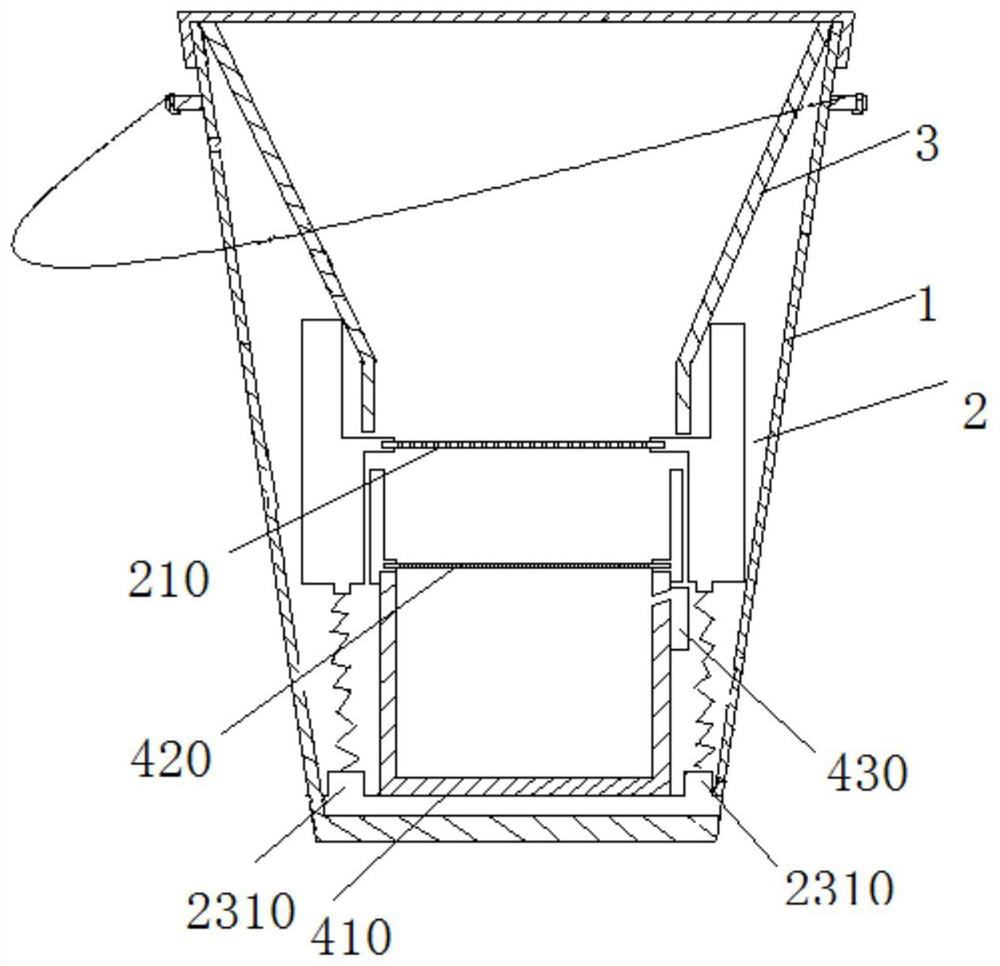
Malignant pleural effusion tumor cell separation device and method

A technology for malignant pleural effusion and tumor cells is applied in the field of malignant pleural effusion tumor cell separation device, which can solve the problems of low filter porosity and inability to complete a large-capacity clinical fluid enrichment process.

Pending Publication Date: 2021-05-18
PEKING UNIV FIRST HOSPITAL
View PDF0 Cites 0 Cited by
  • Summary
  • Abstract
  • Description
  • Claims
  • Application Information

AI Technical Summary

Problems solved by technology

However, the porosity of these filters is still very low, and can only achieve a filtration flux of less than 2mL / min, which cannot complete the enrichment process of large-capacity clinical fluids.

Method used


Image

  • Malignant pleural effusion tumor cell separation device and method
  • Malignant pleural effusion tumor cell separation device and method
  • Malignant pleural effusion tumor cell separation device and method

Examples


Embodiment 1

[0058] The positive detection rate of tumor cells was compared between the filtering device in Example 1 and the centrifugal smear method.

[0059] 4. Test results

[0060] 1. 39 cases of pleural effusion specimens were studied and found that the filter membrane separation device and separation method of the present embodiment 1, the detection rate of tumor cells exfoliated by bronchoalveolar lavage fluid (BALF) was increased to 80%, compared with Compared with the traditional cytocentrifugation method, the detection rate is only 45%. The specific data are as follows: figure 2 As shown in (a), the separation result picture is as follows image 3 shown.

[0061] 2. In addition, the filter membrane separation device and separation method of the present embodiment 1 have greatly improved the detection sensitivity compared with the traditional cell centrifugation method, and the specific data are as follows: figure 2 (b) shown.

[0062] 3. Compared with the filter membrane s...


PUM


Abstract

The invention provides a malignant pleural effusion tumor cell separation device and method. The separation device comprises a containing tank, a vibration filter sieve, a drainage funnel and a filter cup, wherein the vibration filter sieve, the drainage funnel and the filter cup are all located in the containing tank; the vibration filter sieve is provided with a first microfiltration membrane; and the filter cup is provided with a second microfiltration membrane. The filter cup is arranged in the vibration filter sieve, so that the second microfiltration membrane is aligned with the first microfiltration membrane. The micropore diameter of the first microfiltration membrane and the micropore diameter of the second microfiltration membrane are set, so that thymic tumor cells can remain on the second microfiltration membrane when pleural effusion containing cells to be removed sequentially passes through the first microfiltration membrane and the second microfiltration membrane. The separation method comprises a use method of the separation device. The separation device and method have the advantages that the tumor cells can be quickly and efficiently separated and enriched from the pleural effusion, a high-purity and high-concentration sample can be provided for detection and identification of the downstream pleural tumor cells, the pathogen detection time is shortened, and the pathogen detection accuracy is improved.

Description

technical field [0001] This application relates to the field of advanced manufacturing and processing and biomedical technology, in particular to a malignant pleural effusion tumor cell separation device and separation method. Background technique [0002] Malignant pleural effusion (Malignantpleural effusion, MPE) refers to the primary malignant tumor in the pleura or other parts of the malignant tumor metastasis to the pleural effusion caused by. At present, there is still a lack of investigation and research data on the epidemiology of MPE in China. According to statistics, the annual incidence of MPE in the United States exceeds 150,000. Corrected by the population base, it is estimated that about 1,200,000 new cases of malignant pleural effusion occur in my country every year. MPE can occur in almost all malignant tumors. Lung cancer is the most common cause, accounting for about 1 / 3 of MPE, followed by breast cancer. MPE is not uncommon in lymphoma, ovarian cancer, an...

Claims


Application Information

Patent Timeline
IPC IPC(8): C12M1/12C12M1/00
CPCC12M47/04
Inventor 林连君魏珊辰张继新刘姚萍王玮刘新民
Owner PEKING UNIV FIRST HOSPITAL
Learn More