Mouse lung organ culture method

A technology of organ culture and organoids, applied in the field of cell biology, can solve the problems of low formation rate of lung organoids, difficult manipulation and cumbersome culture process, etc., shorten the in vitro culture period, shorten the culture period, and simplify the experimental steps Effect

Active Publication Date: 2021-05-28
NANJING MEDICAL UNIV
View PDF4 Cites 2 Cited by
  • Summary
  • Abstract
  • Description
  • Claims
  • Application Information

AI Technical Summary

Problems solved by technology

[0005] In prior art methods, lung organoids are mainly derived from embryonic stem cells or induced pluripotent stem cells, and their development requires activation of relevant signal transduction in a controllable manner at an appropriate time pathway, induce cell fate differentiation and spatial separation to form different cell types, and guide self-organization formation (stem cells generate endoderm, which then forms three-dimensional tissues), but the manipulation of this microenvironment is not easy to control, and the culture process is cumbersome and time-consuming , low rate of lung organoid formation, poor activity, and large variability in size, structural organization, function, and gene expression

Method used


Image

  • Mouse lung organ culture method
  • Mouse lung organ culture method
  • Mouse lung organ culture method

Examples


Embodiment 1

[0052] Firstly, adopt the method shown in CN111534477A to cultivate the primary epithelial stem cell spheres of mouse lung tissue (that is, the following steps 1 to 10).

[0053] (1) Thoroughly disinfect the skin of the mouse with 75% alcohol, aseptically separate the lung tissue and rinse it in pre-cooled sterile PBS (phosphate buffered saline solution), remove the connective tissue and the main bronchus in the lung, and separate the lung lobes at the same time, Wash 2-3 times to remove blood.

[0054] (2) Transfer the cleaned lung lobe to a new cell culture dish with sterile tweezers, suck off the residual PBS, and use ophthalmic surgical scissors to evenly cut the lung tissue into about 1mm 3 The tissue pieces were transferred to the preheated collagenase digestion solution and digested on a constant temperature shaker (100 rpm) at 37°C for 45-60 minutes;

[0055] (3) Add an equivalent amount of DMEM / F12 medium containing 10% FBS to stop the digestion, pipette and mix well, ...

Embodiment 2

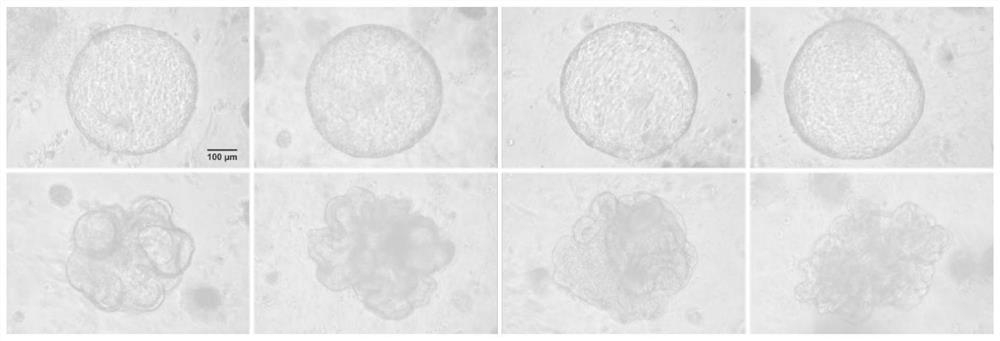
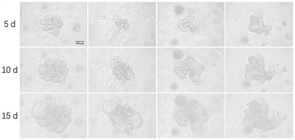
[0075] In this example, the effect of lung spheroids with different diameters on organoid culture was tested.

[0076] Such as Figure 6 As shown, the lung spheroids Figure 6 Left); lung spheroids > 80 μm were used for subsequent organoid culture, and organoids with obvious hollow and branched structures could not be formed ( Figure 6 right). Only when the lung spheres of 50-80 μm are selected, can the hollow and circular alveolar organoids and lung bud organoids that spontaneously realize the morphological development and differentiation of the lungs be formed.


PUM


Abstract

The invention discloses a mouse lung organ culture method, which comprises the following steps: using 50-80 [mu] m mouse lung tissue primary epithelial stem cell spheres for mouse lung organ culture, adding matrigel according to the number of lung spheres, incubating a mixture of the lung spheres and Matrigel basement membrane matrigel to cure the matrigel, and culturing by using a lung organ culture medium. Two different forms of lung organs can be simultaneously differentiated and formed under the same culture system.

Description

technical field [0001] The invention belongs to the technical field of cell biology, in particular to a method for culturing mouse lung organoids. Background technique [0002] At present, monolayer adherent cell culture on two-dimensional (2D) substrates (such as polystyrene or glass) has become the main research method of traditional cell culture systems, but the morphological characteristics, growth patterns, and physiological functions of cells in 2D culture There are obvious differences with the real environment in the body, and the physiological correlation is not strong. In recent years, the development of a large number of 3D cell culture models has significantly promoted the research and development of tumor biology, tissue engineering, and regenerative medicine. The 3D cell culture model can self-assemble into microspheres through suspension culture or induce differentiation to form organoids based on biological scaffolds (such as extracellular matrix). Cell-cell ...

Claims


Application Information

Patent Timeline
Patent Type & Authority Applications(China)
IPC IPC(8): C12N5/071
CPCC12N5/0688
Inventor 周宏孔辉解卫平曾晓宁黄文刘萍
Owner NANJING MEDICAL UNIV
Why Eureka
Try Eureka