A method for detecting aflatoxin b1 based on fluorescent copper nanoparticles

An aflatoxin and nanoparticle technology, which is applied in the fields of analytical chemistry, materials science, and nano-biosensing to achieve highly sensitive fluorescence detection and high selectivity.

Active Publication Date: 2022-04-29
JIANGNAN UNIV
View PDF11 Cites 0 Cited by
  • Summary
  • Abstract
  • Description
  • Claims
  • Application Information

AI Technical Summary

Problems solved by technology

[0006] Food safety regulations are increasingly strict on the residue limit of AFB1 in food. Using simple fluorescent nanoprobe design to achieve high sensitivity, high selectivity, and high accuracy detection, there is no relevant research report so far

Method used


Image

  • A method for detecting aflatoxin b1 based on fluorescent copper nanoparticles
  • A method for detecting aflatoxin b1 based on fluorescent copper nanoparticles
  • A method for detecting aflatoxin b1 based on fluorescent copper nanoparticles

Examples


Embodiment 1

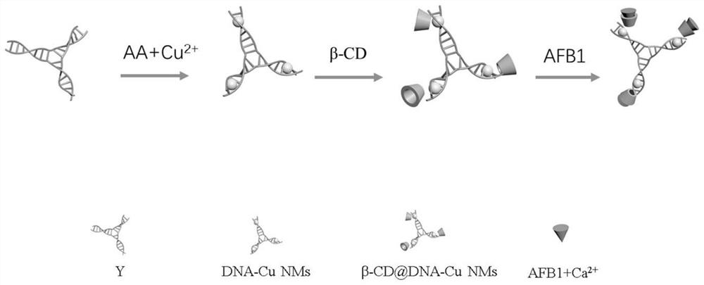
[0073] A method for preparing fluorescent copper nanoparticles β-CD@DNA-Cu NMs, comprising the steps of:

[0074] (1) Preparation of template strand Y-type complementary nucleic acid duplex (Y-type DNA):

[0075] Three equal oligonucleotide strands (Y 0a ,Y 0b ,Y 0c ) (Table 1) in MOPS buffer (10mM, pH 7.5, 150mMNaAc, 1mM MgCl 2 ) to obtain a mixed solution; then, the mixed solution was heated to 90°C, denatured for 5 minutes, and then slowly cooled to room temperature to obtain Y-shaped DNA; the Y-shaped DNA solution was stored in a -18°C refrigerator.

[0076] Table 1 DNA sequence of oligonucleotide chain

[0077] serial number sequence (from 5' to 3')) Y 0a

CCTGTCTGCCTAATGTGCGTCGTAAG SEQ ID NO.1 Y 0b

CTTACGACGCACAAGGAGATCATGAG SEQ ID NO.2 Y 0c

CTCATGATCTCCTTTAGGCAGACAGG SEQ ID NO.3

[0078] (2) Preparation of DNA-Cu NMs:

[0079] Mix Y-shaped DNA solution (500 μL, 2 μM) with ascorbic acid solution (500 μL, 1.25 mM), t...

Embodiment 2

[0083] The optimization of embodiment 2 fluorescent copper nanoparticles preparation technology

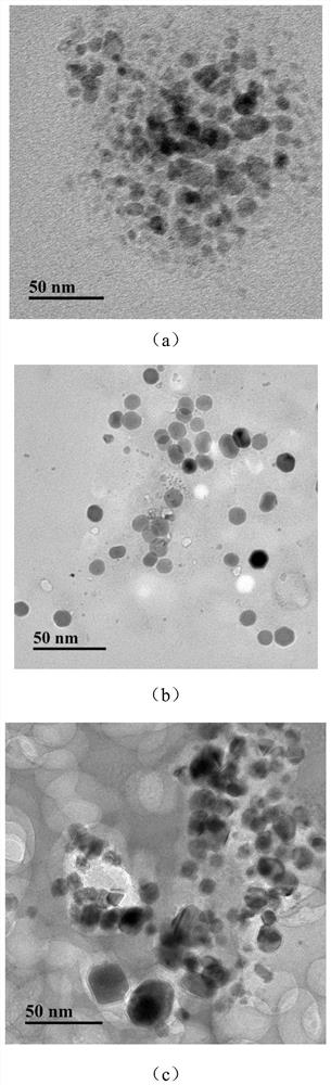
[0084] The concentration of the β-CD solution in Example 1 was adjusted to 0.5, 2, and 5 mM, and the others were kept the same as in Example 1 to obtain β-CD@DNA-Cu NMs.

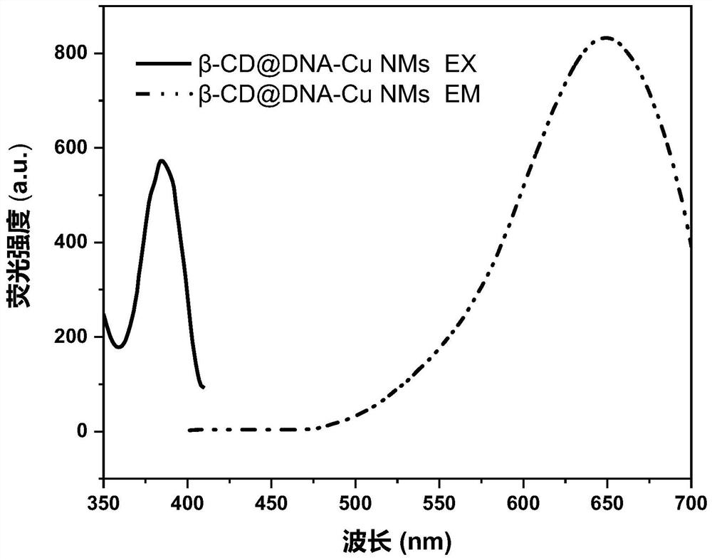
[0085] The obtained β-CD@DNA-Cu NMs were subjected to a fluorescence test. The conditions of the fluorescence test were: the excitation broadband was 10nm, the slit width was 10nm, and the excitation wavelength was 365nm; the test results were as follows:

[0086] figure 2 are the TEM images of β-CD@DNA-Cu NMs modified with different concentrations of β-CD; where (a) is 0.5mM, (b) is 2mM, and (c) is 5mM. From figure 2 It can be seen that as the concentration of β-CD increases from 0.5 to 2 and 5 mM, the average size of β-CD@DNA-Cu NMs increases from 10 nm to 12 nm and 14 nm, respectively. The concentration of β-CD is in the range of 0-5mM. With the increase of its concentration, the fluorescence is stronger...

Embodiment 3

[0089] A method for preparing a ratiometric fluorescence sensor of β-CD@DNA-Cu NMs-AFB1, comprising the steps of:

[0090] Add the β-CD@DNA-Cu NMs solution (100 μL, 200 μM) obtained in Example 1 into the AFB1 solution (900 μL, 10 ppb), shake at 400 rpm for 1 min, and mix well to obtain β-CD@DNA-Cu NMs-AFB1 ratiometric fluorescent sensor.

[0091] The obtained β-CD@DNA-Cu NMs-AFB1 ratiometric fluorescence sensor was tested for fluorescence. The test conditions were: the excitation broadband was 10nm, the slit width was 10nm, the excitation wavelength was 365nm, the detection was 433nm (AFB1) and 650nm (β- CD@DNA-Cu NMs) fluorescence values ​​of the two emission peaks.


PUM


Abstract

The invention discloses a method for detecting aflatoxin B1 based on fluorescent copper nanoparticles, and belongs to the technical fields of analytical chemistry, material science and nanobiological sensing. The present invention uses Y-shaped DNA as a template, ascorbic acid as a reducing agent, and β-CD as a fluorescence stabilization enhancer to prepare β-CD@DNA-Cu NMs; then a ratiometric fluorescence detector is constructed with β-CD@DNA-Cu NMs Needle; Finally, the detection of AFB1 with high sensitivity, high selectivity and high accuracy was realized by using fluorescent probe. When the method of the present invention is 0.03-10ppb and 10-18ppb in the linear range, I 433nm / I 650nm The ratio of AFB1 and the concentration of AFB1 show a good linear relationship, the detection limit is 0.012ppb (S / N=3); metal ions can be replaced by Yb 3+ , Y 3+ 、Er 3+ 、Pt 2+ The same applies to improving the detection limit of AFB1 in rice.

Description

technical field [0001] The invention relates to a method for detecting aflatoxin B1 based on fluorescent copper nanoparticles, and belongs to the technical fields of analytical chemistry, materials science and nanobiological sensing. Background technique [0002] Aflatoxin (AFT) is one of the most common pollutants in grains. It is a kind of mycotoxin widely distributed in nature and has strong teratogenicity, carcinogenicity and physiological and metabolic toxicity. Among them, aflatoxin B1 (AFB1) is the most toxic and carcinogenic, and is classified as a class I carcinogen by the International Agency for Research on Cancer (IARC). Long-term consumption of foods containing low levels of aflatoxin will cause the liver to accumulate toxicity, causing liver damage and liver cancer, which seriously threaten human health and even life. Traditional detection methods are mainly instrumental analysis methods such as high-performance liquid chromatography (HPLC), liquid chromatogra...

Claims


Application Information

Patent Timeline
Patent Type & Authority Patents(China)
IPC IPC(8): G01N21/64
CPCG01N21/6402G01N21/6486
Inventor 彭池方李敏邰胜梅魏新林
Owner JIANGNAN UNIV
Why Eureka
Try Eureka