Tryptophan optical probe as well as preparation method and application thereof
An optical probe, tryptophan technology, applied in the field of optical probes, can solve the problems of inability to monitor the change of tryptophan concentration in living cells in real time, complicated steps, and prone to human errors.
- Summary
- Abstract
- Description
- Claims
- Application Information
AI Technical Summary
Problems solved by technology
Method used
Image
Examples
Embodiment 1
[0139] Example 1: Tryptophan-binding protein particles
[0140] The 3lft gene in the Streptococcus pneumoniae gene was amplified by PCR, and the PCR product was recovered after gel electrophoresis and digested with HindIII and XhoI, and the pCDF vector was subjected to corresponding double digestion. After ligation with T4 DNA ligase, the product was used to transform DH5α, and the transformed DH5α was spread on LB plates (streptomycin 100ug / mL), and cultured at 37°C overnight. The growing DH5α transformant was extracted from the plasmid and then identified by PCR. After the positive plasmid is correctly sequenced, the subsequent plasmid construction is carried out.
Embodiment 2
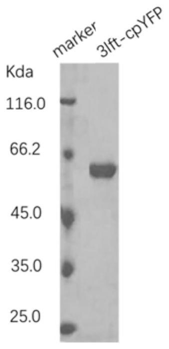
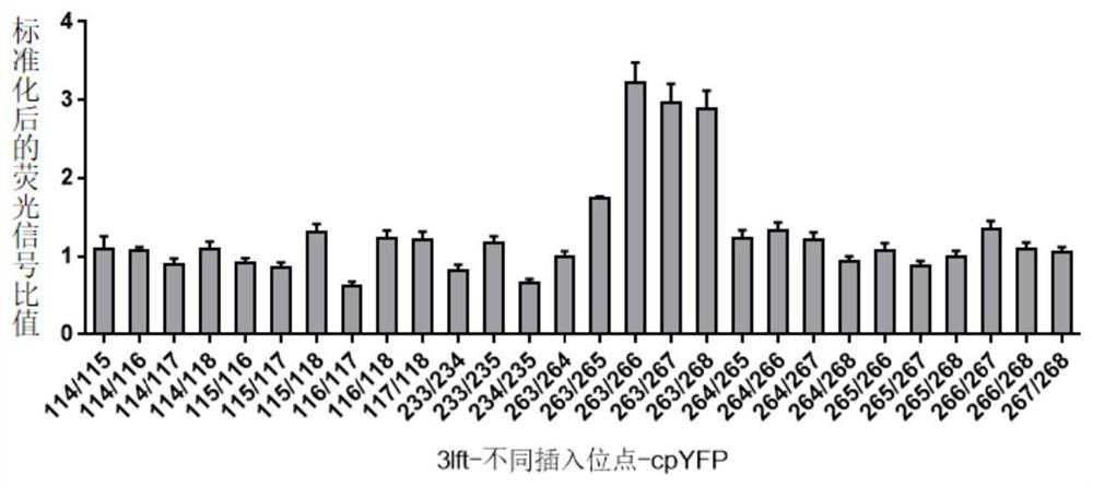
[0141] Example 2: Expression and detection of cpYFP optical probes at different insertion sites
[0142] In this example, based on pCDF-3lft, the following sites were selected to insert cpYFP according to the crystal structure of tryptophan-binding protein, and the corresponding pCDF-3lft-cpYFP plasmids were obtained: 114 / 115, 114 / 116, 114 / 117, 114 / 118, 115 / 116, 115 / 117, 115 / 118, 116 / 117, 116 / 118, 117 / 118, 233 / 234, 233 / 235, 234 / 235, 263 / 264, 263 / 265, 263 / 266 , 263 / 267, 263 / 268, 264 / 265, 264 / 266, 264 / 267, 264 / 268, 265 / 266, 265 / 267, 265 / 268, 266 / 267, 266 / 268 or 267 / 268. Exemplary sequences are shown in Table 1.
[0143] Table 1. Sequences of Optical Probes
[0144] sequence insertion site SEQ ID NO:6 263 / 265 SEQ ID NO:7 263 / 266 SEQ ID NO:8 263 / 267 SEQ ID NO:9 263 / 268
[0145] The cpYFP DNA fragment was generated by PCR, and the cpYFP terminal homologous sequence was introduced through the 5' end of the primer, and the pCDF-tryptophan b...
Embodiment 3
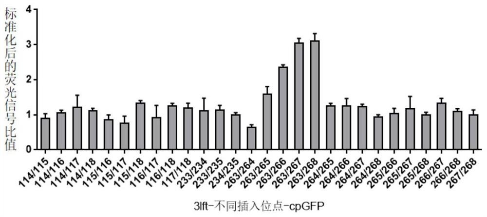
[0148] Example 3: Expression and detection of cpGFP optical probes at different insertion sites
[0149] According to the method in Example 2, cpYFP was replaced with cpGFP to construct a tryptophan green fluorescent protein fluorescent probe. Such as image 3 As shown, the detection results of the broken supernatant show that the optical probes that respond to tryptophan more than 1.5 times are implemented at the 263 / 265, 263 / 266, 263 / 267 and 263 / 268 positions or the corresponding amino acid positions of their family proteins Insert the optical probe.
PUM
 Abstract
Description
Claims
Application Information
 - R&D
- Intellectual Property
- Life Sciences
- Materials
- Tech Scout
- Unparalleled Data Quality
- Higher Quality Content
- 60% Fewer Hallucinations
Browse by: Latest US Patents, China's latest patents, Technical Efficacy Thesaurus, Application Domain, Technology Topic, Popular Technical Reports.
© 2025 PatSnap. All rights reserved.Legal|Privacy policy|Modern Slavery Act Transparency Statement|Sitemap|About US| Contact US: help@patsnap.com


