Microelectrode-based tumor marker rapid high-sensitivity detection method

A tumor marker, sensitive detection technology, applied in the field of microelectrode detection, can solve problems such as applications that have not yet been reported, and achieve the effects of high sensitivity, strong affinity and low cost

Pending Publication Date: 2021-10-08
BINZHOU MEDICAL COLLEGE +1
View PDF0 Cites 2 Cited by
  • Summary
  • Abstract
  • Description
  • Claims
  • Application Information

AI Technical Summary

Problems solved by technology

[0019] However, the application of aptamer-based microelectrodes

Method used


Image

  • Microelectrode-based tumor marker rapid high-sensitivity detection method
  • Microelectrode-based tumor marker rapid high-sensitivity detection method
  • Microelectrode-based tumor marker rapid high-sensitivity detection method

Examples


Embodiment Construction

[0057] The present invention will be specifically introduced below in conjunction with the accompanying drawings and specific embodiments.

[0058] 1. Detection of tumor marker CEA based on microelectrodes

[0059] Step 1. Making platinum microelectrodes

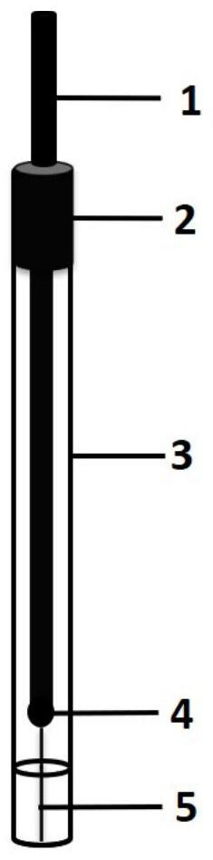
[0060] refer to figure 1 First, connect one end of the platinum wire 5 to one end of the copper wire 1 through graphite-filled conductive glue 4, then insert the platinum wire 5 downwards and the copper wire 1 upwards into the capillary glass tube 3 as a whole, and make the end of the platinum wire 5 Flush with the lower end of the capillary glass tube 3, make the end of the copper wire 1 exceed the upper end of the capillary glass tube 3, then seal the upper end (electrode copper wire end) of the capillary glass tube 3 with epoxy resin glue 2, and dry at room temperature. The capillary glass tube 3 is fused with the platinum wire 5 by a flame melting method, and finally the lower end of the capillary glass tube 3 (the end...


PUM

PropertyMeasurementUnit
Diameteraaaaaaaaaa

Abstract

The invention discloses a microelectrode-based tumor marker rapid high-sensitivity detection method, wherein a tumor marker is a CEA. The detection method comprises the following steps of synthesizing a CEA aptamer and modifying the tail end of the CEA aptamer with sulfydryl, manufacturing a bare electrode, depositing gold nanoparticles on the surface of the bare electrode, then combining the gold nanoparticles with the sulfydryl-modified CEA aptamer, drawing a standard curve, detecting a clinical sample, and determining the content of CEA in a sample according to the standard curve. The method has the beneficial effects that (1) the microelectrode is used as a current type sensor, so that the rapid and high-sensitivity detection of the tumor marker in a small-volume serum sample can be realized; (2) the aptamer is used as a specific recognition molecule, the synthesis is simple, the stability is good, the affinity is strong, and the selectivity is high; and (3) electrochemical detection based on square wave voltammetry is low in cost, is simple and convenient to operate and is rapid in detection.

Description

technical field [0001] The invention relates to a tumor marker detection method, in particular to a microelectrode-based rapid and highly sensitive detection method for tumor markers, belonging to the technical field of microelectrode detection. Background technique [0002] Tumor markers can stably exist in body fluids or tissues such as serum and urine, and can indicate the existence and growth changes of tumors, and are important indicators for early diagnosis of tumors. [0003] The realization of rapid and highly sensitive detection of tumor markers has always been the main requirement of clinical detection. [0004] At present, the detection methods of tumor markers include: radioimmunoassay, chemiluminescence immunoassay, enzyme-linked immunosorbent assay, immunosensor, proteomics, molecular biology method, liquid biopsy, electrochemical sensor, etc. [0005] 1. Radioimmunoassay [0006] As a traditional tumor marker detection method, radioimmunoassay has the advant...

Claims


Application Information

Patent Timeline
IPC IPC(8): G01N27/327G01N27/48
CPCG01N27/3277G01N27/3278G01N27/48
Inventor 赵光涛刘凯翟佳丽
Owner BINZHOU MEDICAL COLLEGE
Why Eureka
Try Eureka