CIK cell culture solution and culture method for enhancing CD3+CD56 + of CIK cell
A culture method and cell culture technology, applied in the field of enhancing CIK cell CD3+CD56+ culture, can solve the problems of difficult to meet clinical needs, low activation efficiency, poor proliferation effect of CIK cells, etc.
- Summary
- Abstract
- Description
- Claims
- Application Information
AI Technical Summary
Problems solved by technology
Method used
Image
Examples
preparation example Construction
[0030] 1. Preparation of peripheral blood mononuclear cells
[0031] 1.1. Take 50-60ml of peripheral blood from healthy people, collect it in a heparin bottle, and store it at 37°C.
[0032] 1.2. Use two 50ml centrifuge tubes (marked 1 # and 2 # ) into 20ml of lymphatic separation fluid.
[0033] 1.3. Use a ground glass pipette to transfer 30ml of peripheral blood to two centrifuge tubes filled with lymphocyte separation medium (note: the peripheral blood needs to be placed on the upper layer of lymphocytes).
[0034] 1.4. Place the two centrifuge tubes in a centrifuge and centrifuge at 800g for 15min at 20°C.
[0035] 1.5. After centrifugation, use a negative pressure device to suck as much plasma as possible from the two centrifuge tubes, and transfer the resulting lymphocyte layer cells to another 50ml centrifuge tube (marked 3 # )middle.
[0036] 1.6. According to the dosage of 80,000 units of gentamicin in 500ml of normal saline, add 160IU / ml of gentamicin in normal ...
Embodiment 1
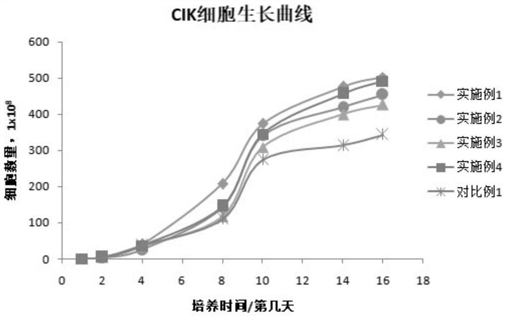
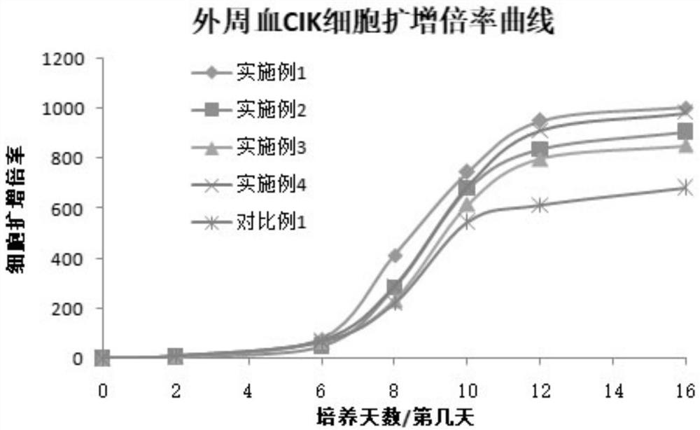
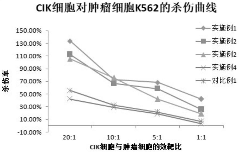
[0056] Cells were removed from the above liquid nitrogen at a concentration of 0.5 × 10 8 Peripheral blood mononuclear cells (PBMC) per ml were quickly placed in a water bath at 37°C-42°C, shaken quickly to melt within 1 minute; the outer surface of the cryotube was disinfected with alcohol, and moved to a biological safety cabinet; Transfer the cells in the cryopreservation tube to a 50ml centrifuge tube in a biological safety cabinet; centrifuge at 1500rpm for 10min, 4°C, increase speed to max, and decelerate to max; Resuspend in RPMI-1640 medium with % autologous serum, adjust the cell concentration to 1×10 after counting 6 within the range of cells / ml, and then inoculated into T175 culture flasks.
[0057] On the first day, add RPMI-1640 medium containing 10% autologous serum in a volume of 25 ml to the culture flask, then add IFN-2γ cytokine, and control the final concentration of IFN-2γ cytokine in the medium to 1000 μg / ml; then put the culture bottle into the incubat...
Embodiment 2
[0062] Cells were removed from the above liquid nitrogen at a concentration of 0.5 × 10 8 Peripheral blood mononuclear cells (PBMC) per ml were quickly placed in a water bath at 37°C-42°C, shaken quickly to melt within 1 minute; the outer surface of the cryotube was disinfected with alcohol, and moved to a biological safety cabinet; Transfer the cells in a cryopreservation tube to a 50ml centrifuge tube in a biological safety cabinet; centrifuge at 1500rpm for 10min, 4°C, increase speed to max, and decelerate to max; after centrifugation, absorb the supernatant in the centrifuge tube, and use Resuspend in RPMI-1640 medium with 10% autologous serum, adjust the cell concentration to 1×10 after counting 6 within the range of cells / ml, and then inoculated into T175 culture flasks.
[0063] Add IFN-2γ cytokines to the culture flask, and control the final concentration of IFN-2γ cytokines in the medium to 500 μg / ml, add RPMI-1640 medium containing 10% autologous serum to a volume o...
PUM
 Abstract
Description
Claims
Application Information
 - R&D
- Intellectual Property
- Life Sciences
- Materials
- Tech Scout
- Unparalleled Data Quality
- Higher Quality Content
- 60% Fewer Hallucinations
Browse by: Latest US Patents, China's latest patents, Technical Efficacy Thesaurus, Application Domain, Technology Topic, Popular Technical Reports.
© 2025 PatSnap. All rights reserved.Legal|Privacy policy|Modern Slavery Act Transparency Statement|Sitemap|About US| Contact US: help@patsnap.com


