Automatic mitral valve ring displacement detection system based on ultrasonic image

An automatic detection and ultrasound image technology, applied in the field of computer vision, can solve the problems of not being able to meet the clinical needs, the cost of learning time, and the dependence of evaluation results, so as to reduce the cost of learning and time, speed up the calculation, and avoid the results. effect of difference

Pending Publication Date: 2021-11-02
NORTHEASTERN UNIV +1
View PDF0 Cites 0 Cited by
  • Summary
  • Abstract
  • Description
  • Claims
  • Application Information

AI Technical Summary

Problems solved by technology

However, this method requires the doctor to manually trace three sites in the software, and the evaluation results depend on the doctor's clinical experience and the accuracy of the tracing position, and the repeatability is poor
Manual tracing requires investment in learning, labor and time costs, and cannot well meet clinical needs

Method used


Image

  • Automatic mitral valve ring displacement detection system based on ultrasonic image
  • Automatic mitral valve ring displacement detection system based on ultrasonic image
  • Automatic mitral valve ring displacement detection system based on ultrasonic image

Examples


Embodiment Construction

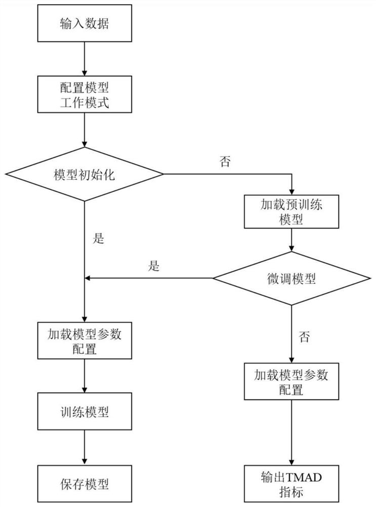
[0041] The specific implementation manners of the present invention will be further described in detail below in conjunction with the accompanying drawings and embodiments. The following examples are used to illustrate the present invention, but are not intended to limit the scope of the present invention.

[0042] In this embodiment, a mitral annulus displacement automatic detection system based on ultrasound images, through such as figure 1 The following steps are shown for the detection of mitral annulus displacement:

[0043] Step 1: Acquire multiple echocardiograms as a sample dataset; based on the echocardiogram Figure four Cardiac view, to obtain echocardiogram files in dcm format following the Digital Imaging and Communications in Medicine (DICOM), or single-frame images (JPG, PNG, JPEG) after parsing echocardiogram files in dcm format format) and its corresponding left ventricular mask or mitral annulus site coordinate label;

[0044] Step 2: Divide the sample dat...


PUM


Abstract

The invention provides an automatic mitral valve ring displacement detection system based on an ultrasonic image, and relates to the technical field of computer vision. According to the system, firstly, a plurality of echocardiograms with left ventricle mask or mitral valve ring site coordinate labels are obtained to serve as a sample data set; then a deep neural network model for left ventricle segmentation and mitral valve detection is initialized, and the deep neural network model is pre-trained by using the sample data set to obtain a pre-trained model; model parameters and a configuration file of the pre-training model are loaded, a left ventricular intima in the echocardiogram to be evaluated is segmented, and a mitral valve ring ventricular septum site coordinate, a left ventricular side wall site coordinate and a left ventricular apex site coordinate are determined; and mitral valve ring ventricular septum site displacement, left ventricle side wall site displacement and displacement variation of the mitral valve ring relative to the cardiac apex from the initial systole stage to the final systole stage are calculated, and an MAD index is output on the to-be-evaluated echocardiography image to complete automatic detection of the mitral valve ring displacement of the echocardiography image.

Description

technical field [0001] The invention relates to the technical field of computer vision, in particular to an automatic detection system for mitral annulus displacement based on ultrasonic images. Background technique [0002] Left ventricular systolic function is an important parameter for evaluating cardiovascular diseases. The first choice for evaluating left ventricular systolic function clinically is left ventricular ejection fraction (Left Ventricular Ejection Fraction, or LVEF). In the early stage of some diseases, LVEF cannot accurately reflect the reduction of cardiac systolic function. The overall longitudinal strain of the ventricle (Left Ventricular Global Longitudinal Strain, LVGLS) was analyzed and evaluated. However, this method needs to trace the endocardium of the left ventricle, and the accuracy of assessment is highly dependent on image quality. Mitral Annular Displacement (MAD) is another index to evaluate left ventricular systolic function, which tracks ...

Claims


Application Information

Patent Timeline
Patent Type & Authority Applications(China)
IPC IPC(8): G06T7/00G06T7/10G06T7/73G06N3/04G06N3/08
CPCG06T7/0012G06T7/10G06T7/73G06N3/08G06T2207/10132G06T2207/30048G06N3/045
Inventor 杨金柱马春燕李洪赫瞿明军王永槐曹鹏冯朝路覃文军栗伟
Owner NORTHEASTERN UNIV
Why Eureka
Try Eureka